
Download PDF published by SfN
(Chapter from 'The History of Neuroscience in Autobiography, Volume 8' edited by Larry R. Squire)
Yuh-Nung Jan and Lily Jan
Birth
Family History and Growing Up
National Taiwan University
The Hiking Trip to Shitou in the Spring of 1967
Graduate School Application
Graduate Study at Caltech (1968–1974)
Seymour Benzer Lab (1974–1977)
Steve Kuffler’s Lab at Harvard Medical School and Life in Boston and Woods Hole (1977-1979)
University of California, San Francisco (1979–present)
Breakthroughs in 1987
Our Family and Life Outside of the Lab
Some Reflections
References
Lily and Yuh-Nung Jan went to Caltech in 1968 after their undergraduate study in physics at National Taiwan University. After two years of graduate study in physics, they switched to biology under the influence of Max Delbrück. They stayed at Caltech for postdoctoral training with Seymour Benzer and then worked in Steve Kuffler’s lab at Harvard Medical School to demonstrate that peptides can function as neurotransmitters. During their postdoctoral training with Seymour Benzer, they began their long-term collaboration. Cloning of the first potassium channel gene Shaker and its mammalian homolog in the Jan lab at University of California, San Francisco (UCSF), followed up with expression cloning of a founding member of the inwardly rectifying potassium channels and the founding member of a novel calcium-activated chloride channel family, has led to molecular and cell biological studies of how these ion channels work and how they contribute to neuronal signaling. In parallel to these ion channel studies, the Jans started their work on neural development at UCSF in order to understand how neurons acquire their specific cell fate and morphology. Their discoveries include atonal—a founding member of the large family of proneural genes that endow cells with neuronal cell fates, and numb—the first cell fate determinant exhibiting asymmetric localization in dividing neural precursor cells. More recently, they have begun unraveling the logic and underlying mechanisms for generating diversity in neuronal morphology (especially dendritic morphology) and learning how such diversity contributes to the wiring of the nervous system
Yuh-Nung Jan
Born:
Shanghai, China
December 20, 1946
Education:
National Taiwan University, BS (1967)
California Institute of Technology, PhD (1974)
Appointments:
Postdoctoral Research Fellow, California Institute of Technology (1974)
Postdoctoral Research Fellow, Harvard Medical School (1977)
Assistant Professor, University of California, San Francisco (1979)
Investigator, Howard Hughes Medical Institute (1984)
Honors and Awards (Selected):
McKnight Scholar Award (1978–1981)
Elected member, National Academy of Sciences (1996)
Elected member, Academia Sinica, Taiwan (1998)
Distinguished Alumni Award, California Institute of Technology (2006)
Elected member, American Academy of Arts and Sciences (2007)
Javits Neuroscience Investigator Award, National Institute of Neurological Disorders and Stroke,
National Institutes of Health (2010)
Seymour Benzer Lecture, Neurobiology of Drosophila meeting, Cold Spring Harbor Lab (2011)
Honors and Awards Shared by Lily Jan and Yuh-Nung Jan:
W. Alden Spencer Award and Lectureship, Columbia University (1988)
38th Faculty Lecturer Award, University of California, San Francisco (1995)
Harvey Lecture, New York (1998)
The Stephen W. Kuffler Lecture, Harvard Medical School (1999)
K. S. Cole Award, Biophysical Society (2004)
Jan Lab Symposium (2006)
Society of Chinese Bioscientists in America Presidential Award (2006)
Ralph Gerard Prize, Society for Neuroscience (2009)
Edward M. Scolnick Prize in Neuroscience, Massachusetts Institute of Technology (2010)
Albert and Ellen Grass Lecture, Society for Neuroscience (2010)
Wiley Prize in Biomedical Sciences (2011)
Gruber Neuroscience Prize (2012)
Lily Jan
Born:
Fu-Chow, China
January 20, 1947
Education:
National Taiwan University, BS (1968)
California Institute of Technology, PhD (1974)
Appointments:
Postdoctoral Research Fellow, California Institute of Technology (1974)
Postdoctoral Research Fellow, Harvard Medical School (1977)
Assistant Professor, University of California, San Francisco (1979)
Investigator, Howard Hughes Medical Institute (1984)
Honors and Awards (Selected):
Alfred P. Sloan Research Fellowship (1977–1979)–
Klingenstein Fellowship Award (1983–1986)
Elected member, National Academy of Sciences (1996)
Elected member, Academia Sinica, Taiwan (1998)
Distinguished Alumni Award, California Institute of Technology (2006)
National Institute of Mental Health MERIT Award (2006)
Elected member, American Academy of Arts and Sciences (2007)
Honors and Awards Shared by Lily Jan and Yuh-Nung Jan:
W. Alden Spencer Award and Lectureship, Columbia University (1988)
38th Faculty Lecturer Award, University of California, San Francisco (1995)
Harvey Lecture, New York (1998)
The Stephen W. Kuffler Lecture, Harvard Medical School (1999)
K. S. Cole Award, Biophysical Society (2004)
Jan Lab Symposium (2006)
Society of Chinese Bioscientists in America Presidential Award (2006)
Ralph Gerard Prize, Society for Neuroscience (2009)
Edward M. Scolnick Prize in Neuroscience, Massachusetts Institute of Technology (2010)
Albert and Ellen Grass Lecture, Society for Neuroscience (2010)
Wiley Prize in Biomedical Sciences (2011)
Gruber Neuroscience Prize (2012)
Yuh-Nung Jan and Lily Jan
Birth
We were born nine days apart. Yuh-Nung’s birthday was listed as December 20, 1946, but that is according to the lunar calendar commonly used then. It corresponds to January 11, 1947, in the Western calendar, which is nine days ahead of Lily’s birthday, January 20, 1947. Because Yuh-Nung was born slightly ahead of his due date, we might have started our embryonic development around the same time.
Yuh-Nung’s father, Ten-Sun Jan, was from a well-to-do family in Jiangxi Province, China. Originally, Yuh-Nung’s grandfather planned to send his son to the United States to study economics. However, the outbreak of the Sino-Japanese war in 1937 altered the plan. Instead, Yuh-Nung’s father finished college in China and then joined the Nationalist army to resist Japanese invasion. He became an officer during World War II. Yuh-Nung’s mother, Li-Ju Chen, was from Anhui Province and went to the same college as his father. They got married toward the end of the war and moved to Shanghai. Yuh-Nung was born there as their first son.
In 1949, as communists were taking over, Yuh-Nung’s parents got out of mainland China at the last minute and escaped to Taiwan. During the first few years in Taiwan, the family lived in a small rural town of Xinpu in Hsin-Chu County, about 70 km south of Taipei. At the time, Yuh-Nung’s father was still in the military and was stationed in the garrison in the frontline (Quemoy Island). His mother had to work and needed childcare. She persuaded the local elementary school to take in Yuh-Nung as a first-grader on a trial basis even though he was only four and a half years old. Yuh-Nung was able to keep up academically with the older kids in the class, so the school let him continue. Later, after his father left the military and started working for the government, the family moved to Taipei, and Yuh-Nung entered the Jian-Guo High School, which was (and still is) the top high school for boys in Taipei (the counterpart of Lily’s Taipei First Girls’ High School).
Initially, Yuh-Nung was an indifferent student at the Jian-Guo High School. He spent most of his time reading novels, daydreaming, playing sports and the game of go, and so on and paid little attention to schoolwork. He earned barely passable grades. In his junior year, something clicked inside him. Influenced by two superb science teachers (in biology and chemistry) and several academically strong classmates that were his good friends, he became very interested in science, especially physics and chemistry. In the last year of high school, he became very motivated and studied hard in preparation for attending a university to study science.
At the time, the university admission system in Taiwan had some unusual features and was quite different from that in the United States. The students were admitted into a particular university department, so each student had to decide on their major before entering college. There were two ways of being admitted to the university and the department of choice. Each of the 10 or so top high schools throughout Taiwan, including Jian-Guo High School and Taipei First Girls’ High School, could send a few students with the highest grades (the top 1 or 2 percent in those schools) directly to the universities. (Lily was one of those very few.) The vast majority of the students took a common entrance exam for all the universities at the same time. Each student, in a single rank order determined by the results of that exam, was assigned a department in a university based on his or her own priority list; if the slots were filled for the top choice on the list, the department the student would join would be determined collectively by the priority lists and by the entrance exam scores of all high school seniors. At that time, National Taiwan University (NTU) was “the” university among a dozen or so in Taiwan. Acceptance there was very competitive, especially for the most desirable departments—such as physics, medicine, electrical engineering, and chemistry—because they would fill their slots very quickly with the topscoring students. In this system, except for the few students such as Lily who could bypass the entrance exam because of stellar high school grades, grades had no bearing whatsoever on university admission. It depended entirely on the results of this intense, annual, two-day exam. Yuh-Nung benefited from this system. Despite his so-so grades, he could choose any department he wanted because he did very well on that exam. (His score was among the top 10 out of approximately 30,000 students.) He agonized over his final two choices—the department of physics and the departmentof medicine at NTU. He could not decide and resorted to a coin toss. The coin toss was for the department of medicine, but he picked the department of physics instead. Eventually, however, he ended up with a career in the school of medicine at University of California, San Francisco (UCSF). That coin toss was “deterministic” after all. Like any traditional Chinese family, Yuh-Nung’s parents cared very much about their kids’ education and future but nevertheless allowed him complete freedom to choose his path. (Several years later, Yuh-Nung’s only sibling, his younger brother, Jonathan, followed his footsteps into the physics department of NTU.)
To go back a bit, Lily was born shortly after the end of World War II in Fu-chow, China. Her parents, Hong-Shu Yeh and Chuan-Hwa Lee, both accountants, brought her to Taiwan as a baby. There, she grew up and was drawn into science while attending Taipei First Girls’ High School, a fairly large school with several dozen students in each of a score of classrooms for the six grades. All students had neatly trimmed short hair that was not allowed to extend past the earlobe, and all wore the unusual uniform of green shirts, black skirts, white socks, and canvas shoes. A subset of students was assigned to Principal Jiang’s “experimental classrooms”—the Kung (fairness), Chung (sincerity), Qing (diligence), and Yi (perseverance) classroom (Ban in Chinese). Lily was in the Qing Ban and studied subjects such as three-dimensional geometry. Lily was fortunate to have a chemistry teacher who offered students opportunities such as reading college level textbooks and working on additional problem sets that he took the time to grade and for which he provided feedback. So, when faced with the choice of three tracks for college entrance, Lily chose the track for math and sciences. When given the opportunity to choose a department, Lily knew very little about the different departments and made her choice more capriciously than by tossing a coin. When told by Lily’s high school classmate of her elder brother’s advice about the two departments not suitable for girls—physics and electrical engineering—Lily then chose the physics department.
Around that time, students in Taiwan were influenced by the awarding of the 1957 Nobel Prize in physics to Tsung Dao Lee and Chen Ning Yang for their theoretical work on nonconservation of parity in weak interactions. Chien-Shiung Wu, the experimental physicist who demonstrated this parity violation, made multiple visits to Taiwan to talk to young students in the 1960s. Lily has vivid memories of Wu’s lectures and her advice to aspiring science students to consider biophysics as an emerging field of interest. Not quite following Wu’s advice, Lily chose to pursue graduate study in theoretical high-energy physics when she was an undergraduate student in the physics department of National Taiwan University (NTU)
Yuh-Nung entered the physics department of NTU in 1963, and Lily entered the same department the following year. At that time, it was mostly a teaching rather than a research-oriented department. The research facility and the opportunity to be mentored by top-notch faculty were nowhere near physics department standards for major research universities in the United States (although it is much better now). However, the students were an outstanding group because the department was among the hardest to get in. In general, students were smart and very motivated, and they formed study groups and inspired one another. Although lacking the experience in cutting edge research, these students did receive a strong foundation in physics and mathematics, which prepared them well for pursuing advanced training, mostly in U.S. graduate schools. Many of our classmates went on to have successful careers in a variety of fields. Perhaps the most notable is Andrew Chi-Chih Yao. Andrew and Yuh-Nung had lived on the same street in Taipei (their families lived three houses apart) since their early teens. They were classmates, friends, and rivals in high school and at the university. Andrew was exceptionally good at mathematics. After earning his PhD in physics, he switched to computer science. He was a professor at Stanford and then at Princeton. In 2000, he received the Turing Award, the highest honor in computer science. In 2004, he moved to China to direct the Institute for Theoretical Computer Science at Tsinghua University in Beijing.
The Hiking Trip to Shitou in the Spring of 1967
Because more than 90 percent of the physics students were male and had mandatory military service the year after college, Lily was with the class one year ahead of hers when applying for graduate school, and she went along for their graduation trip as well. At that time, traveling within the island of Taiwan was still a big deal reserved for special occasions such as the grand field trip celebrating graduation from college. The destination of that year’s graduation trip was Shitou, a beautiful forest recreation area located in the mountainous region of central Taiwan. During that week of train rides and hiking in the mountains, we got to know each other. Yuh-Nung was smitten and began pursuing Lily. Many years later, we participated not only in one special Wu Chien-Shiung Science Camp but also several Wu Ta You Science Camps. The latter were week-long camps organized by Academia Sinica in honor of Wu Ta You, a distinguished physicist who was a former Academia Sinica president and a mentor to Tsung Dao Lee and Chen Ning Yang. For this camp, around a hundred or so bright undergraduates (selected from Taiwan and several countries in Eastern Asia) who were interested in biomedical or physical sciences spent the week with a number of experienced scientists and were exposed, in alternating years, to various areas of modern biology or physics. We participated not only because it was a worthy cause but also for sentimental reasons because the camps were held in Shitou, where we first met.
During Lily’s senior year in NTU, Yuh-Nung fulfilled his military service as a communication and electronics officer in the Taiwanese Air Force stationed at an air base about 30 km from Taipei. He got a motorcycle so he could ride to Taipei to see Lily every chance he could sneak out of the base. One of the things we were doing together was applying for graduate school.
Nowadays, one can pursue advanced studies in Taiwan. Back then, one had to go abroad (mostly to the United States). Because we both wanted to do theoretical high-energy physics, Caltech was our dream school. Its physics department had the towering figures of Richard Feynman and Murray Gell-Mann as well as a constellation of stellar theoretical and experimental physicists. In prior years, very few NTU physics students were admitted into the Caltech physics department. As luck would have it, in 1967, the department accepted a student, Wei-Dou Ni, from the NTU physics department in the class one year ahead of Yuh-Nung’s. Wei-Dou performed superbly at Caltech and that undoubtedly helped the subsequent applications of NTU physics students. In 1968, the Caltech physics department accepted three students, Chi-Shin Wang (a brilliant student in Yuh-Nung’s class) and the two of us from NTU; this was unprecedented.
Chi-Shin became a very successful entrepreneur in Silicon Valley. After Caltech, Chi-Shin got a degree in electrical engineering at Stanford. After working for Hewlett Packard for a few years, he started his own company in Silicon Valley. Among his successes were his pioneering commercial applications of the global positioning system (GPS).
Graduate Study at Caltech (1968–1974)
Physics Department (1968–1970)
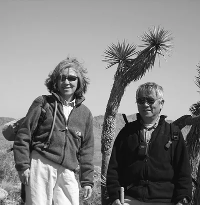
In September 1968, we arrived in Pasadena from Taipei (see Figure 1). For this first trip overseas, we brought all our belongings in two suitcases that could be carried. Caltech is an academic institution with a superb student to faculty ratio of about four to one and a very small but highly selective student body. In 1968, most of the 750 undergrads and 750 graduate students had nice housing on campus. Yuh-Nung stayed in one of the buildings for male graduate students. However, with no female undergraduate students and only a very few female graduate students, in 1968, the Caltech faculty had just created a graduate women’s house. In the corner house at 293 South Chester Street, Caltech converted seven rooms into bedrooms and there Lily joined six other female graduate students. She stayed in this brand new dorm for three years. To satisfy the fire code for a dorm, which required multiple fire exits for the two upstairs bedrooms, Lily was handed some ropes and told to throw the ropes out of her bedroom window in case of a fire. These ropes were only used once—for a demonstration; somehow nothing ever triggered the fire alarm even with the seven students taking turns cooking dinner every week.
Switch to Biology (1970)
As fellow graduate students in theoretical physics, we became aware of the excitement exuded by biology graduate students we ran into in the dorms and in the small Caltech community. Although we went to Caltech for its great physics department, it happened also to have a fabulous biology division that gave us our first exposure to modern biology. What triggered the switch from physics to biology was a speaker invited by Max Delbrück for the weekly physics seminar in 1970 who introduced the basic concepts of molecular biology and enzymology, hoping to entice physics students to consider doing research in biology. When Yuh-Nung went to talk to Max afterward, Max thought that he wanted to join his lab, and Yuh-Nung thought why not? He returned with a small flowerpot with a stalk of the fungus Phycomyces growing in it and became a graduate student in Max’s lab. Now that Yuh-Nung had joined Max’s lab, Lily thought she would find a different lab for her thesis study, so she knocked on the office doors of the rest of the biology faculty and asked them to please tell her what their research was about. What Lily recalls most vividly was the visit with Jerome Vinograd, an outstanding biophysicist. Instead of telling Lily about his research, he offered the advice that if she wanted to switch from physics to biology, “Don’t try to apply physics to biology; do what Max Delbrück did by becoming a biologist and thinking like a biologist.” When Lily joined Max’s membrane biology subgroup, she began her apprenticeship by painting lipid bilayers for recording the currents generated by carrier molecules and got to work in a smog-free room in the sub-basement of the electrical engineering building. From then on, Max made sure that there was a total separation of Lily’s graduate study from Yuh-Nung’s pursuit of the sensory transduction processes, including the mysterious avoidance response of Phycomyces.
In 1971, we were married. It was the time of the Vietnam War, Woodstock, and general social upheaval in United States. We felt no inclination or obligation to have a traditional wedding, so we chose the simplest ceremony possible. It cost just six dollars to get a marriage license and pay for parking at the Los Angeles courthouse; the mandatory blood tests were free for students. Two witnesses were needed. With foresight, we asked three friends to drive to the courthouse to witness the ceremony (in case one got caught in the Los Angeles traffic). Two made it through the traffic in time to serve as witnesses when a judge married us in his chamber. The next day, we celebrated by camping and hiking at Yosemite. We moved out of the graduate student housing and settled in a little detached cottage on Euclid Street near Caltech. The rent was only $70 a month.
Having gone through the grueling qualifying exams for physics graduate students before switching to biology and then barely passing the placement test on organism biology (so the Caltech biology department did not have to offer this elementary course for the first time to someone utterly unprepared but who somehow turned up in the entering class), we joined fellow biology graduate students in a rebellious challenge to the seven-day, open-book, open-library written exam customarily administered to students at the end of their first year. The faculty patiently worked with our class in multiple meetings to find an alternative qualifying exam format that was mutually agreeable, and they accepted our suggestion that each student write a research proposal and then be tested in an oral exam in which the student would defend his or her own proposal plus a fellow student’s proposal. This original format may have been implemented only for the qualifying exams of our class (students in subsequent years probably thought we were nuts and came up with their own ideas—perhaps more reminiscent of the major and minor proposals).
For this experimental qualifying exam, Lily came up with her own idea for a proposal to localize rhodopsin in photoreceptors and decided to stick to this proposal for her thesis study. At a time when raising antibodies for immunostaining with electron microscopy was a new experimental approach that entailed purification of not only the protein to be used for immunizing the rabbit but also the antibody along with the ferritin or hemocyanin to be conjugated to the antibody, Lily asked Jean Paul Revel, who had just moved from Harvard to Caltech, to join Max Delbrück as her co-mentors. Then she promptly disappeared into either Max’s darkroom in the basement or into Jean Paul’s darkroom in the sub-basement for the dissection of chicken eyes and purification of rhodopsin according to her naïve plan. Yuh-Nung helped out by repeatedly driving Lily to the chicken slaughterhouse to collect chicken heads in a giant ice chest because Lily did not learn to drive until much later. Lily’s fellow graduate students also offered help with their expertise in rabbit immunization. Somehow the rabbit receiving rhodopsin injections could tell Lily was nervous and would thump his hind foot to show his displeasure when he heard her footsteps in the hallway. Most unfortunately, shortly after the last boosting shot of chicken rhodopsin, the rabbit displayed the ultimate displeasure by dying of anaphylactic shock. Despite Jean Paul Revel’s valiant efforts to salvage some usable antibody from this rabbit, all those long hours laboring in the darkroom for the isolation of photoreceptor outer segments from thousands of chicken eyes and the ensuing rhodopsin purification over the course of one year came to naught, at a time when Lily was trying to figure out whether she was cut out for the career path of an experimentalist in biology. The blessing in disguise turned out to be her switch from chicken eyes to cow eyes as the source for rhodopsin; rabbit antibody against cow rhodopsin worked nicely for localizing rhodopsin in mouse photoreceptors—on the densely packed discs within as well as on the plasma membrane (Jan and Revel, 1974).
For Yuh-Nung’s thesis, he worked on two projects concerning sensory transduction processes using the fungus Phycomyces. The sporangiophore of Phycomyces is a giant single cell. It is cylindrical in shape and can reach several cm in length. It was chosen by Max to study sensory responses because it can sense light and gravity. Phycomyces displays a mysterious “avoidance response.” Its growing zone, a segment of the cell several mm long near the tip of the stalk, can sense the presence of any object placed a few mm away and grow away from it. This happens in the darkness, and it does not matter what the object is made of. Yuh-Nung tried hard to figure out what signal is sensed by Phycomyces, and he was able to rule out just about anything he could think of. For a while, he and his coworkers thought that it sensed wind current (Cohen et al., 1975), but that was ruled out several years later. To this day, the nature of this “avoidance response” remains a mystery.
The main part of Yuh-Nung’s thesis was his attempt to begin to understand the molecular mechanism underlying the sensory transduction processes of Phycomyces. Because Phycomyces responds to various stimuli by altering the elongation rate of its cell wall, which is composed of chitin fibers, Yuh-Nung reasoned that the enzyme chitin synthetase could be an important readout. He characterized this enzyme biochemically and indeed showed that blue light regulates the activity of this enzyme (Jan, 1974).
Max Delbrück, Our PhD Thesis Advisor
We were extremely lucky to have Max as our thesis advisor. With his unique combination of intellect, moral integrity, and charisma, Max was a marvelous mentor and a great influence on us during our particularly impressionable stage. One piece of advice from Max was, “Don’t do fashionable science.” Max liked to venture into unchartered research areas and detested entering a field because it was popular or trendy, a trait shared by our other mentors, Seymour Benzer and Steve Kuffler. Of course, venturing into terra incognito carries risks. In Max’s case, his later choice of Phycomyces to understand sensory transduction was not so successful because although Phycomyces has the advantage of being a large single cell that has a rich repertoire of sensory responses, unfortunately, it is an organism not well suited for genetics or biochemistry. However, his earlier choice of phage to study the basis of heredity was a great success that helped launch modern molecular biology.
The virtue of not following the crowd is nicely articulated in this passage from the biography Genius about Richard Feynman (Gleick, 1992):
“It will not do you any harm to think in an original fashion.” Feynman said. He offered a probabilistic argument. “The odds that your theory will be in fact right, and that the general thing that everybody’s working on will be wrong, is low. But the odds that you, Little Boy Schmidt, will be the guy who figures a thing out, is not smaller. . . . It’s very important that we do not all follow the same fashion. Because it is ninety percent sure that the answer lies over there, where Gell-Mann is working, what happens if it doesn’t?”
“If you give more money to theoretical physics,” he added, “it doesn’t do any good if it just increases the number of guys following the comet head. So it is necessary to increase the amount of variety . . . and the only way to do it is to implore you few guys to take a risk with your lives that you will never be heard of again, and go off in the wild blue yonder and see if you can figure it out.”
Max not only influenced our scientific outlook but also exposed us to many other things that enriched our lives through regular camping trips and through gatherings in Delbrück’s house as part of an extended scientific family—for example, camping under the stars in the desert or reading the works of Samuel Beckett, whose writings Max was very fond of. Max was delighted that the year he was awarded the Nobel Prize in Physiology or Medicine with Luria and Hersey, the literature laureate was Samuel Beckett.
Accepted into Seymour Benzer’s Lab for Postdoc (1974)
In 1973, the thesis work for both of us began to take shape, and it looked like we might get our PhD degrees in another year or so; it was time for us to start figuring out what to do next for postdoctoral training.
One day, Yuh-Nung came upon a paper by Yoshiki Hotta and Seymour Benzer, “Mapping of behavior in Drosophila mosaics” published in Nature (Hotta and Benzer, 1972), in which Hotta and Benzer showed that by making genetic mosaics and constructing embryonic “fate maps” it was possible to locate the anatomical site of abnormalities affecting behavior. It was a very elegant and interesting paper. It got us interested in the ongoing work in Benzer’s lab, and we became very attracted to his approach and to his lab. Benzer was very picky about accepting people into his lab. Fortunately, with some persuasion by Max Delbrück, Seymour accepted us.
We then tried to get postdoctoral fellowships to support our work in Benzer’s lab, with Yuh-Nung proposing to look for learning mutants and Lily proposing to fine-tune mosaic fate mapping of the neural circuitry for visual responses. Unfortunately, the National Institutes of Health (NIH) deemed that neither was worthy of an NIH postdoctoral fellowship so we had to look for less conventional funding sources. Lily somehow got the endorsement of an ophthalmologist from UCLA for her application for a Fight-for-Sight fellowship. For Yuh-Nung, Benzer said: “Why don’t you apply to this Scottish Rite Schizophrenia Research Program fellowship?” because Seymour firmly believed that one could use a fly to study just about any problem in biology including schizophrenia; meanwhile, Yuh-Nung was thinking, “Gee, schizophrenia? Fly? How am I going to pull this off?’ This was back in the era before cloning (BC) and preceding the realization of the remarkable conservation of mechanisms underlying many biological processes. After a while, an idea emerged—there is a fly mutant called tan, which has an abnormal electroretinogram (ERG). It is as if the fly is seeing things and displaying ERG signals when there is nothing to see, and visual hallucination is a hallmark of schizophrenia. Moreover, tan mutants have abnormally low levels of dopamine, and abnormalities in biogenic amines were thought of as a potential cause of schizophrenia.
With a giant leap of faith, Yuh-Nung wrote a proposal including this sentence: “The existence of a link between catecholamine abnormality and a visual defect that is analogous to hallucination in Drosophila mutant tan suggests that it might be used as a model system for schizophrenia.” He sent it in and thought they probably would just laugh at the proposal. To his astonishment, they actually funded his application. His disbelief soon morphed into delusion: “I can’t believe they bought this, imagine what I can do with a less outlandish proposal, maybe I have a future in this business.” Shortly after, the bubble burst when Yuh-Nung attended a small neuroscience meeting. A very prominent neuroscientist saw his nametag and told him: “Ah, I read your proposal. I don’t believe a word of it, but since you are working with Seymour, you’ll do alright.”
Cold Spring Harbor Summer Courses and the Beginning of Our Collaboration (1974)
To prepare for our postdoc in the field of neuroscience, we took one lecture course followed by a lab course in the summer of 1974 at Cold Spring Harbor (CSH) Laboratory, where we had spent the bulk of our summertime as Max’s graduate students, with Yuh-Nung involved in the Phycomyces workshop every summer and Lily doing lab work and taking various summer courses in that secluded idyllic commune for scientists. Little did we know that those summer courses in neuroscience would have such a big impact on our research direction even before we began our postdoctoral study in Seymour’s lab. Seymour’s graduate student Bill Harris was attending the same summer courses with us, and for the one-week break between courses, the three of us visited Woods Hole and also visited Doug Kankel, who did his postdoc with Seymour before joining the Yale faculty. On our way out of Doug’s lab, Bill picked up a bottle of fruit flies and handed it to us, so we could get acquainted with our future experimental animal. That is exactly what we did for the last three days of the lab course.
Although the lecture course was taught by Mike Dennis, Regis Kelly, Carla Shatz, and Eric Frank, all hailing from Harvard’s neurobiology department—the only department in neuroscience at that time—the lab course instructors came from all over the world and included Jac Sue Kehoe and Phillip Ascher (from France) and Enrico Stefani (at that time from Mexico). In that era before the invention of patch-clamp recording, we learned to record from Aplysia neurons and frog muscles. For the last three days of free period left to the students to try whatever they would like, we dug out Drosophila larvae from the mushy food in that old bottle to try out what we had just learned to do with the frog neuromuscular junction, and we enjoyed much encouragement and tutoring from our instructors. It turns out the larval muscles are comparable to frog muscle fibers in diameter but much shorter, so the muscle response to nerve stimulation or iontophoresis of the transmitter glutamate could be readily recorded (with our beginner’s luck) during this lab exercise.
Those CSH summer courses were a fantastic way to learn neurobiology quickly. We not only learned a great deal but also made friends with some great people—our teachers and fellow students. One of our fellow students that we got to know very well was Bob Horvitz. He was on his way to start his postdoc with Sydney Brenner at the medical research council (MRC) Cambridge. He would later make tremendous contributions in developmental biology and especially in programmed cell death, for which he was awarded a Nobel Prize in 2002. Those CSH summer courses were very intense. We worked 16 hours non-stop every day and were exhausted by the end of week two of those three-week-long courses. Bob is the only person we know who took three CSH courses in a row, an early indication that he would have a great career with his tremendous energy and intellect.
Seymour Benzer Lab (1974–1977)
Identifying Shaker as a Likely Potassium Channel Gene
In August 1974, we started in Seymour’s lab. Yuh-Nung began by working on learning and memory as part of the team that did the initial dunce work (Dudai et al., 1976). After spending a few months training flies, we thought that, in order to get to the mechanisms of learning and memory, we needed to develop some functional assay for synaptic transmission, for example with electrophysiology. So, we teamed up to put together an electrophysiology rig in Seymour’s lab in the fall of 1974 to continue what we started in that summer at CSH to characterize larval neuromuscular junction (NMJ) and develop it as an assay for a genetic dissection of synaptic transmission. At that time, the Journal of Physiology was considered the top journal for neurobiology, and it was acceptable for us to take the time and circulate our manuscripts among friends and our CSH summer course instructors until we felt it was ready for the journal. We took pride in publishing our first collaborative work (Jan and Jan, 1976a, b).
Assuming we could go through the recording of a couple of preparations in a few hours, we developed a routine for screening the larvae of behavioral mutants in Seymour’s collection to look for abnormal neuromuscular transmission. Very soon after we started this project, on April 28, 1975, we recorded from a male ShakerKS133 mutant larva and found it displayed an extremely large “unit size” for the excitatory junctional potential in low calcium Ringer—whereas normally each nerve stimulation induced either no response (a failure) or a unit response of the same size as the miniature excitatory junctional potential. Stimulation of the motor nerve of this Shaker mutant generated large responses every time (see Figure 2). In the summer of 1975, the CSH symposium happened to feature the synapse. After Seymour gave a talk and showed our findings based on recordings using a dissecting microscope. Mike Dennis, who taught us in the lecture course the previous summer, offered to teach us to do recordings using a compound microscope with Nomarski optics that would allow us to see the nerve terminals and to do extracellular recordings to test whether the mutant phenotype could be attributed to the nerve or to the muscle of Shaker mutant larvae. We happily made multiple drives to San Francisco. Each time, we stayed for a couple weeks in the Mariana’s guesthouse across the street from the UCSF campus on Parnassus so that we could spend as much time as possible in Mike’s lab in the physiology department. Working closely with Mike, we could see that the Shaker mutant nerve responded to a single stimulation with multiple recurrent action potentials so that calcium iontophoresis several milliseconds after the nerve stimulation could still induce transmitter release and muscle response.

At this point, we wrote up a paper with Mike with the conclusion that the abnormality of the Shaker mutant nerve terminal could be a defect in the potassium channel or in the calcium channel and submitted it to Nature. While this paper was under review, we did more experiments and found that the Shaker mutant phenotype could be phenocopied by applying the potassium channel blocker 4-aminopyridine to wild-type larval preparations. When the Nature editor informed us that the reviewers’ comments were favorable but that we needed to shorten our manuscript significantly, we told the editor that with these additional experiments we would have to lengthen our paper instead. So we withdrew it from Nature and submitted our paper to Proceedings of Royal Society because Seymour was just elected as a foreign member, and we thought he would get a kick out of communicating our paper to the Royal Society journal (Jan et al., 1977). Again, the reviewers’ comments were favorable, and this time one reviewer asked the editor to reveal his identity—Sir Bernard Katz—and to pass on a number of follow-up questions that he was curious about. After we did a series of experiments to address these interesting questions, we wrote a long letter to Katz and included those results in that letter. Though we never got around to publishing those studies, we included one set of results in the 1997 review article in Journal of Physiology (Jan and Jan, 1997).
While we worked with Mike Dennis on Shaker mutant recordings, his colleague John Heuser was working very hard next door looking for some physical evidence of exocytosis at the motor nerve ending. Using a fancy machine custom-made at UCSF to strap a frog nerve muscle preparation around a suspended piston, which was triggered to slam onto a cold slab chilled with liquid helium shortly after the delivery of a nerve stimulus, John would recover that flattened frog muscle for freeze fracture and then disappear into the electron microscope room to search for omega-shaped contours at the end plate that had to be very close to the muscle surface to have been frozen immediately on impact with the cold slab. In one of those neighborly chats with Mike and John, we wondered whether the Shaker mutant larvae with prolonged transmitter release could make the task easier to accomplish. When it became evident that the geometry of the larval nerve terminal—a large bouton rather than the nicely elongated end plate of the frog motor nerve ending—was not amenable to freeze fracture, we turned to the alternative approach of treating the frog nerve-muscle preparation with 4-aminopyridine. Indeed, this treatment also prolonged transmitter release from the frog motor nerve endings making it rather easy to capture vesicle fusions using John’s machine (Heuser et al., 1979). Elated with this success, we had a very memorable celebratory dinner outing with John and Mike at the trendy restaurant Café Sports.
Life in the Benzer Lab
The three years we spent in Seymour’s lab were a wonderful experience—tremendously enjoyable and intellectually stimulating. Seymour had a great sense of humor and an immense curiosity. He was a really fun and influential person to be with. He attracted a very talented and somewhat eccentric group of people—fellow postdocs Chip Quinn, Alain Ghysen, Ilan Deak, and Yadin Dudai, and Seymour’s graduate students Bill Harris, Don Ready, and Duncan Byers were there when we joined the group. Several became lifelong friends, especially Alain Ghysen, who had a strong influence on our later scientific direction. Several important lines of research were initiated in Seymour’s lab during that period. For example, Don Ready laid the groundwork for Drosophila eye development (Ready et al., 1976), and Bill Harris discovered the sevenless mutant (Harris et al., 1976), which led to insights about the mechanisms of induction in retinal cell fate specification.
Before we had our first child, toward the end of our stay in Seymour’s lab, we were “owls” with respect to our circadian rhythm. Each weekday, we would get up just before noon and go to Seymour’s cramped lunchroom to eat with the whole group. There were lively and free-flowing conversations and gossip about science, movies, and often food, a favorite subject of Seymour’s. Those lunches could last for hours. Seymour was a good friend of many prominent scientists, and from time to time they joined in those lunches as well. One of the most memorable was the time when Richard Feynman came over from the physics department. He asked us what we were doing with our Drosophila learning studies. In a couple of hours, he managed to think of every clever experiment that had taken several of us months to come up with. His mind was really impressive.
In the afternoon, after those long lunches, we started our daily work or went to seminars/lectures. After dinner, we came back to the lab and often stayed till 2–3 a.m. We could work uninterrupted for six or seven hours and that was when we did most of our experiments. Another night owl was Ed Lewis. He had an even more extreme schedule. He often came back to the lab around midnight and worked till dawn. That was the period when Ed made a breakthrough in his studies of the bithorax complex by using the embryo cuticles to analyze the various chromosome deficiencies and mutants of the bithorax complex; this made it possible to decipher the effect of lethal mutations on body patterning and to expand the analyses beyond previous studies done with adult flies (Lewis, 1978). On several occasions, Ed was very excited by his new findings and wanted to show someone; we were often the only ones around. We got a glimpse of his progress and shared his excitement. Because we were then studying the larval neuromuscular preparation, we even started a little collaboration with Ed to see how the internal tissues might be altered in the bithorax mutants. As with much of Ed’s work, that was never published. Nevertheless, it was a privilege to get to know Ed. Years later, when Ed organized a symposium in honor of Seymour’s 70th birthday in 1991, it was a wonderful opportunity to meet up with Seymour’s old friends and disciples (see Figure 3).
What to Do Next?
Having gone into the field of neurogenetics with the hope that genetics could help with the identification of key genes for neural signaling in much the same way as it did for biosynthetic pathways, we were hopeful that molecular biological approaches being pioneered by David Hogness and colleagues for positional cloning of Drosophila genes might apply to Shaker cloning with the potential of molecular identification of a potassium channel without having to take on the daunting task of purifying potassium channels. However, cloning was in its infancy, and without any training in molecular biology we were not ready to take on such a challenging task. We were wondering what to do next. Mike Dennis had spent some time previously as a postdoc with Steve Kuffler at Harvard Medical School (HMS) and thought that it might be a good idea for us to go there to work with Steve to gain more experience in neurophysiology. At that time, the department of neurobiology at HMS was “the” neurobiology department in the country. Steve was a towering figure in neurobiology, and there were many really outstanding people in that department. Seymour also endorsed this idea because Seymour was a good friend of Steve’s and many other people in that department, and he had a very high regard for them. Around that time, an Ivy League school asked whether we might be interested in applying for a faculty position. We agonized over whether to start applying for faculty jobs or to do a second postdoc at HMS. We spent a long afternoon walking and talking in the beautiful garden of the Huntington Library near Caltech to try to figure out what to do. Finally, we decided that getting additional postdoctoral training with Steve and spending some time in that great department could only benefit us in the long run.
Birth of Emily and the Move to Boston
The last months at Caltech before we moved to Boston were a very hectic time for us. Our daughter Emily was born on August 6, 1977, a couple of weeks ahead of her due date, and we were not quite prepared. On August 5, we were at a group meeting in Seymour’s lab. Lily started feeling contractions and glanced at her watch to time them—they were not exactly spaced with five-minute intervals. As the intervals between contractions got shorter, we were frantically trying to reach the Lamaze instructor for the class we had been taking in preparation for our child’s birth in order to get the course certificate that Yuh-Nung needed in order for him to be allowed in the delivery room to serve as the Lamaze coach. As we were driving to the Kaiser hospital in Hollywood, we were trying to come up with a name for the new baby, one for a boy and one for a girl, because we did not know the sex of the baby. After a long night of labor, a wonderful girl, Emily Huan-Ching Jan, was born early the next morning.
Finally, it was time to leave Pasadena. In September 1977, we packed up and drove across country to Boston with seven-week-old Emily. Pasadena and Caltech were our home for nine years, and we spent our entire twenties there. We had arrived in California very naïve—culturally and scientifically— and we grew up there. Caltech influenced us more than anywhere else. It was a great privilege for us to know the tremendous scientists and human beings Max Delbrück, Seymour Benzer, Ed Lewis, and their wonderful families and many other wonderful people at Caltech. In 2006, we both were given the Distinguished Alumni Award at Caltech, one of our most treasured awards, as if our alma mater was telling us “you kids did all right,” akin to parental approval.
Steve Kuffler’s Lab at Harvard Medical School and Life in Boston and Woods Hole (1977–1979)
Peptidergic Transmission for the Late Slow Excitatory Postsynaptic Potential
In late September 1977, we arrived in Boston to join Steve Kuffler’s lab. We were very fortunate to find a very nice flat near Coolidge Corner in Brookline that was a half-hour walk from the lab. Lily’s mother flew from Pasadena to Boston to live with us and helped take care of Emily during the daytime. We began our new routine of taking shifts to cover the long hours in the lab and to tend to a baby at home in the evening. Lily would set up the bullfrog sympathetic ganglia for recording early in the morning and work together with Yuh-Nung during the day. Yuh-Nung would walk Lily home at dinnertime to relieve her mother of babysitting and then return to the lab to resume the experiments.
At that time, Steve was interested in slow synaptic potentials. Initially, he assigned us to map by electrophysiology the distribution of muscarinic receptors, which mediate the slow excitatory postsynaptic potential that lasts for seconds. It was a useful learning experience but was not that interesting a problem. We became restless after six months and discussed starting a new project with Steve. We were attracted by the mysterious late slow excitatory postsynaptic potential (EPSP) lasting for several minutes. The late slow EPSP, which was initially discovered by Nishi and Koketsu, persisted in the presence of antagonists for nicotinic acetylcholine receptors responsible for the fast EPSP and muscarinic acetylcholine receptors responsible for the slow EPSP. Therefore, some unidentified transmitter other than acetylcholine has to induce the late slow EPSP. With Steve’s blessing, we decided to try to identify the transmitter for the late slow EPSP, a very interesting but highly risky project.
To try to identify this mysterious transmitter, in May 1978, we began to apply all kinds of receptor agonists and antagonists to look for an effect on the late slow EPSP. Besides compounds ordered from Sigma and other companies, we found some small vials of peptides in the freezer that were gifts to Steve from Wylie Vale and Jean Revier at the Salk Institute. Perhaps Steve got these peptides because at the time neuroscientists had begun to suspect that peptides might function as neurotransmitters. To save time, we pooled contents from three vials at a time for a quick survey. One combination produced a moderately encouraging response. Testing them individually identified the culprit as “LHRH.” We then looked up to see what LHRH stood for—lutenizing hormone releasing hormone—and learned about the existence of LHRH analogs as potent agonists and antagonists. When we put those analogs on and observed that the agonist could induce responses that mimic the late slow EPSP, and the potent antagonist could block both the LHRH-induced slow depolarization and the nerve-evoked late slow EPSP, we knew that we were probably on the right track in proposing that an LHRH-like peptide could be the transmitter mediating the late slow EPSP. With the new finding, the obvious next step was to demonstrate that an LHRH-like peptide was indeed present in the presynaptic nerve that innervates the bullfrog sympathetic ganglia and could be released under conditions that could elicit the late slow EPSP. The method of choice was radioimmunoassay, with which we had no experience. We were then told by our colleagues that there was this new postdoc, Tom Jessell, who had just joined Gerry Fishbach’s lab at HMS and who was an expert because he had worked on substance P for his PhD thesis in the UK. We went to see Tom, and he gave us many helpful hints.
Then it was time for the Kuffler lab to make its customary migration to Woods Hole for the summer. Originally, the plan was for Steve and for us to work primarily on the electrophysiology of the late slow EPSP, but we ended up doing mostly radioimmunoassays. Steve’s lab at Woods Hole had great electrophysiology set-ups but hardly any biochemistry equipment. However, Woods Hole was such a communal place that we were able to rely on the kindness of the neighbors to do the necessary experiments.
On the top floor of the Woods Hole lab building with an ocean view on one side, Steve’s lab had windows overlooking the tennis courts on the backside—surely not a coincidence given that Steve was an enthusiastic tennis player. Beyond the tennis courts was the apartment for us, a wonderful arrangement for our daughter and her grandmother to be close by and for us to slip in some tennis during incubation times for radioimmunoassays to detect the LHRH-like peptide in bullfrog sympathetic ganglia and to document the release of the peptide upon nerve stimulation. We had a wonderful three month stay at Woods Hole and went through the list of criteria that a putative transmitter must satisfy and ticked them off one by one. We also got to celebrate our daughter’s first birthday and go to the beach for swimming and parties at that vibrant and happy community with a long history as the favorite summer retreat for biologists. One of Steve’s visitors was his old friend and coworker Sir Bernard Katz. That was our first chance to meet him. We chatted while pushing Emily in a stroller and watching Steve display his prowess as a tennis player. We were delighted that he remembered reviewing our Shaker manuscript (Jan et al., 1977) and that he liked the paper.
By the time the summer was over and we all moved back to Boston, we had pretty much established that an LHRH-like peptide is the transmitter that mediates the late slow EPSP. It took only about six months since we started the project. It was mostly dumb luck. We wrote up the paper with Steve, and he communicated it to Proceedings of the National Academy of Sciences (PNAS) (Jan et al., 1979). That work most likely helped several schools become interested in us and encouraged us to apply for faculty positions. We felt it was perhaps time for us to get independent positions and to start our own lab.
Although we spent less than two years in Boston, the experience at the HMS was very valuable. The neurobiology department was relatively small but had a very high concentration of terrific neuroscientists. The senior faculty members were Steve and the other founding members of the department, who had all worked with him in their youth—David Hubel, Torsten Wiesel, Ed Furshpan, David Potter, and Ed Kravitz. The junior faculty members were Paul Patterson, Story Landis, Simon Levay, Peter Maclish, Ann Stuart, and John Hildebrand. Each group typically had only a few highly selected postdocs and students. When we arrived in 1977, Lou Reichardt, Carla Shatz, and Josh Sanes had just left. Our contemporaries included among others: Mike Stryker, Bill Harris, Eric Frank, Doju Yoshikami, Larry Marshall, Bob Stickgold, Marge Livingstone, Alison Doupe, Mary Kennedy, Charlie Gilbert, and Terry Sejnowski. It is remarkable how much impact that relatively small group of people has had on neuroscience.
The department then was very tightly knit. Two core activities were especially educational. Because all members of the department could fit into a modest-sized lunchroom, we all had lunch together every day and often had lunch seminars given by visitors. It was a tough crowd. The speakers were constantly interrupted, and there was an element of “who could ask the most critical question” during those seminars. We remembered that during a Christmas party when we were there, “mock awards” were given out at a skit. One was given to the person “who most consistently anticipated the next slide with questioning during those lunch seminars in the previous year.”
One of the best department activities was the “evening meetings.” Every month or two, the department had dinner together, and one group presented their ongoing work. Everyone took those presentations seriously because there was tremendous peer pressure to do well. During those evening meetings, we learned about much of the exciting work going on in the department.
Job Offer at University of California, San Francisco
After a few months of job interviews, we had several nice job offers and one was from UCSF. At that time UCSF was not yet a prominent place (like it is now), and we had better offers in terms of space and start-up funding from more prestigious places. At UCSF, we had to share a faculty teaching equivalent (FTE). The lab space and the setup money were very modest as stated in the job offer letter written by the chair of the physiology department, Fran Ganong, on December 6, 1978: “ Zach Hall and I have combined our resources to provide 1,000 square feet of space.” (That was for two of us.) And then he wrote: “I can also commit $15,000 start-up money for each of you at this time” ($30,000 total). Although a dollar in 1978 is worth about three dollars now, $30,000 is not a lot of start-up money. Nevertheless, we were very attracted to UCSF, especially by the people there. We already knew Mike Dennis and John Heuser from our previous collaborations. Zach Hall was recruited from HMS to UCSF to start the neuroscience program in 1976, and in 1977 and 1978, he recruited Lou Reichardt and Mike Stryker (both star postdocs) from the neurobiology department at HMS. Additionally, several other young faculty were already there including Roger Nicoll, Regis Kelly, Howard Field, Mike Merzenich, and Allan Basbaum. These young faculty members formed a core that soon developed into one of the country’s leading neuroscience programs. Our other job possibilities were also at excellent places, and there is no way to know how things might have turned out if we went to one of those instead. Nevertheless, we felt very fortunate to have decided to come to UCSF to join this group, and this was one of the best decisions we have ever made.
An unexpected benefit of coming to UCSF that we were not aware of initially was that, between 1976 and 1979, in parallel to the developing neuroscience program, a fabulous biochemistry and biophysics department was being greatly enhanced with the recruitments of these few years: Bruce Alberts, Marc Kirschner, Keith Yamamoto, Christine Guthrie, Pat O’Farrell, and Tom Kornberg. Because molecular biology was beginning to revolutionize neuroscience in the early 1980s, we soon began benefiting from having the opportunity to interact with those outstanding molecular biologists and cell biologists.
University of California, San Francisco (1979–present)
Setting Up Our Modest Little Lab
In late June 1979, we drove across the country during the height of the oil crisis, with our daughter a toddler not quite two years old, to start our lab at UCSF on July 1. It took us a couple of months to set up our little lab on the eighth floor of Health Sciences East (HSE) on the Parnassus campus (see Figure 4) and then we started doing experiments.

Peptide Acting at a Distance
Initially, we continued with our work on peptides as neurotransmitters. We discovered that although the LHRH-like peptide was released together with a classical transmitter, acetylcholine (ACh), from the same nerve terminals that synapse onto the C type neurons in the sympathetic ganglion, the peptide can diffuse over tens of microns to act on their true targets (i.e., nearby B-type neurons with which the LHRH-like peptide containing preganglionic nerve fiber does not form a synapse). So the wiring diagram based on anatomically defined synapses is actually misleading for identifying the real target of the peptide transmitter (Jan and Jan, 1982b; Jan et al., 1980). This is something for the connectome folks to consider.
Starting New Projects: Shaker Cloning and Neural Development Studies (1980)
We might have been recruited to the UCSF faculty as electrophysiologists studying the vertebrate autonomic nervous systems; however, once our peptide work got going, we soon started switching back to studies of the fruit fly Drosophila because of the opportunities offered by new developments in the field. In our youthful exuberance, we initiated two new projects for which we had no relevant expertise whatsoever: neural development and Shaker cloning. Looking back, it seemed rather foolhardy to start such risky projects as beginning assistant professors. Perhaps one reason that we chose to pursue high-risk projects that interested us was because we felt that we had the good will and strong support from our chair, Fran Ganong, and the neuroscience program director, Zach Hall. Indeed, we were tenured rather quickly (in 1983) even though, by that time, our new projects had yielded very little concrete results.
Neural Development
Neural development was a question that had been in the back of our minds for some years. Back in the days when we were in Seymour Benzer’s lab, in one of those long lunch gatherings together with fellow postdocs Alain Ghysen, Ilan Deak, and Yadin Dudai and Seymour’s graduate students Bill Harris, Don Ready, and Duncan Byers, we all unabashedly went to the blackboard in Seymour’s lunchroom to write down the big questions in neuroscience that interested us—a record of those early musings was kept because Seymour took a Polaroid picture of the scribble on the blackboard. How the nervous system forms was one obvious question on the list though it was not clear how one could go about approaching this question.
While in Seymour’s lab, we became very good friends with a fellow postdoc, Alain Ghysen. We thought it would be fun to do some work together at some point. After we set up our little lab at UCSF, Alain and his long-term collaborator Christine Dambly-Chaudiere would come over from their lab in Brussels and work together with us to explore all kinds of crazy ideas for a few weeks at a time every year in the early 1980s. We wanted to work together on something interesting that had not been worked on by any of us already. We chose neural development.
Two papers that appeared around that time suggested an approach to study neural development. The 1980 paper by Nüsslein-Volhard and Wieschaus on “Mutations affecting the segment number and polarity in Drosophila” (Nüsslein-Volhard and Wieschaus, 1980) was inspiring because it revealed the involvement of just a score of genes for the specification of the body plan and demonstrated the power of canvassing the whole genome with comprehensive screens of mutant embryos. Even those “lethal” mutations incompatible with survival can be characterized by examining the cuticle patterns of embryos that may or may not have made it through embryogenesis. The cuticle prep that remained after dissolving away the embryo within retained the body plan signatures for scoring mutations that affect segmentation and polarity but would not work for scoring mutations affecting the nervous system. The hybridoma technology developed by Köhler and Milstein for monoclonal antibody generation (Köhler and Milstein, 1975) offered some hope. Without any knowledge of the molecules involved in neural development, we could simply immunize mice with ground up embryos and screen the monoclonal antibodies based on the staining patterns they yield. That was the plan anyway; and then there were some accidental findings and lucky breaks in this venture.
To get started with monoclonal antibody generation, Sandra Barbel, who was initially planning to join our brand new lab as a technician for a year or two before returning to her graduate study at UC Davis, learned the hybridoma technologies with the helpful advice from Lou Reichardt’s lab. Sandra has remained and is now our lab manager. Yuh-Nung developed a routine of listening to his favorite music while going through hundreds of staining patterns looking for those monoclonal antibodies that appeared to recognize specific cell types or structures, especially ones in the nervous system. This way we began the bootstrap approach of using whatever monoclonal antibodies that emerged from these screens and that could help with the identification of mutant embryos with abnormal distribution of neurons and characterizing those mutants molecularly to end up with more markers for neurons and their precursors that could then be used for additional mutant screens.
Besides the generation and screening for monoclonal antibodies, serendipity smiled several times in our haphazard experiments. Early on, when Yuh-Nung was using horseradish peroxidase (HRP) as a neuronal tracer in the study of the frog autonomic nervous system, he had a vial of antibody against HRP in the refrigerator. Because Lily was doing staining of cryostat sections of fruit flies with antibodies against neuropeptides for no particular reason beyond a simple curiosity, she reached for the vial of secondary antibody and ended up with an amazing staining pattern of the entire nervous system—rather odd and unexpected given the sparse distribution of peptides in the vertebrate nervous system. For the next three days, Lily vehemently argued against Yuh-Nung’s suggestion that she may have made a mistake somewhere, until she repeated the experiment using either the secondary antibody meant for her experiment or the antibody against HRP in the vial on the same shelf. As Yuh-Nung likes to tell the story, because it was Lily’s mistake that led to the surprise finding that antibody against HRP specifically labels Drosophila and grasshopper neuronal membranes, she was the first author of the paper on that study (Jan and Jan, 1982a). While we were going through rounds and rounds of mouse immunization for hybridoma generation, we immunized some mice with HRP for good measure and recovered a monoclonal antibody that prominently marked the germ plasm and germ cells and led Bruce Hay to the molecular characterization of Vasa (Hay et al., 1988). However, this Vasa antibody does not recognize HRP, and we have no explanation other than luck for the way we came up with some of the most useful monoclonal antibodies for our mutant screens.
Even with those neuronal markers, we were still unsure on which part of the nervous system to focus our attention. Around 1985, we finally realized that the larval peripheral nervous system (PNS) is a good assay system for studying neural development. Rolf Bodmer, Alain, Christine, and the two of us worked out an atlas of the larval PNS (Ghysen et al., 1986; Bodmer and Jan, 1987). Then, Alain and Christine made a very critical discovery. They found that mutants of the achaete scute complex (AS-C) displayed a very striking PNS phenotype: one type of sense organ, the external sensory (es) organ, was missing but the chordotonal (cho) organs were not affected (Dambly-Chaudiere and Ghysen, 1987). This was the first, and very nice, example that a mutation can produce a very clear-cut and neuronal type specific phenotype, which encouraged us to plunge into serious genetic screens for mutants affecting PNS development in 1985. That is how genes like numb and atonal were later discovered.
Shaker Cloning
As we got to know Pat O’Farrell well, we would often stop at the hallway and chat when we ran into one another. One day Pat was all excited about the new development with the P-element, the transposon for the mysterious genetic phenomenon known as hybrid dysgenesis. At the time, Pat was collaborating with Tom Kornberg on the chromosome walk to clone engrailed and was raising the possibility of using P-element insertion mutagenesis as a novel approach to clone Shaker. Pat generously allowed us to do a rotation in his lab to learn about molecular biology. For the next several years, there was close collaboration between Diane Papazian, Bruce Tempel, and Tom Schwarz in our lab and Steve DiNardo and Claude Desplan, who joined Pat’s lab as postdoctoral fellows, to go after the Shaker gene. At a time when there were numerous P-elements in a genome prior to the development of reliable control of movement of these transposons, we settled with the old reliable chromosome walk for cloning Shaker. As the years for this chromosome walking, chromosome sitting (we hit an apparently unclonable region), and chromosome falling off (repetitive sequences doing their trick) dragged on and other interesting biological questions such as cell cycle control in the developing Drosophila embryo beckoned, we were left with our three postdocs Diane, Tom, and Bruce to stick with it to the end.
During the long haul for Shaker cloning and genetic screens for neural development mutants, it was also payback time after having taken so many summer courses at Cold Spring Harbor in our graduate student and postdoc years. We revived the neurobiology of Drosophila course and spent four consecutive summers at Cold Spring Harbor, starting in 1984, the year we were expecting the arrival of our son Max. The apartment building for CSH course instructors was up a gentle slope just about a hundred yards from the lab, a wonderful arrangement for us to be close to our kids even with the traditional long hours for the summer courses. Pat joined us in teaching the course for the first year, and some of our postdocs and students came along to help out as teaching assistants. Some of the summer course students decided to do their postdoctoral research with us later on, as in the case of Ehud (Udi) Isacoff. The scientific nickname Udi can be traced to the first day when he arrived at Cold Spring Harbor for the summer course and, while introducing himself to his roommate Claude and struggling with the French pronunciation of Ehud, he ended up with being stuck with his childhood nickname Udi.
Howard Hughes Medical Institute and the Birth of Max (1984)
The year 1984 was an eventful one. Scientifically, we were at a low point. We had been consistently productive ever since our postdoc days (1974–1982), but from 1983 to 1986 we had a dry spell. Shaker cloning was very difficult. Despite three to four years of hard work, we had nothing to show. Feeling in the dark without knowing what a potassium channel should look like, we could not even tell whether we were on track to clone the first potassium channel gene before everything finally clicked in the end. For the neural development work, we were trying to find our way. We had generated some very useful markers and developed the embryonic PNS as a promising system for genetic dissection of neural development, but we had not yet made any substantial inroad.
During this difficult period, two great events happened to us. One was the birth on November 7, 1984, of our son Max Huang-Wen Jan. We named him Max in honor of our PhD advisor Max Delbrück. Following Chinese tradition, all the kids in Yuh-Nung’s extended family of Emily and Max’s generation share a common given name “Huang,” which means “bright” or “brilliant.” For Emily, her specific Chinese given name is “Ching,” which is an endearing form of “person.” For Max, his specific Chinese given name “Wen” means “literature” or “culture.” With Emily and Max, we are blessed with two wonderful kids, each very talented in a different way.
Another great thing that happened to us was that somehow we were chosen as Howard Hughes Medical Institute (HHMI) investigators. At that time HHMI did not yet have an open competition system, they chose their investigators in a somewhat mysterious way. In 1984, HHMI decided to start supporting neuroscience. They picked five institutes, Harvard Medical School, Columbia, Johns Hopkins Medical School, Salk, and UCSF, and asked these institutions to nominate potential investigators. UCSF decided to choose neuroscience faculty who were relatively young and not yet at the rank of full professor. That meant there was a very small pool of potential candidates. Somehow UCSF picked us even though we were struggling mightily to get our research going. HHMI made a huge difference in our research. We are most grateful for their continued support since July 1, 1984, now more than 29 years and counting.
Shaker Cloning (1987) to Enable Studies of Potassium Channels One at a Time
Finally, after six years of hard work, the project of Shaker cloning came to fruition. One day, Diane saw the S4 sequence with the basic residue arginine or lysine at every third position within an otherwise hydrophobic sequence that could pass as a transmembrane segment, so she knew we probably had the real thing. Over the next few months, Diane, Tom, and Bruce isolated a number of alternatively spliced isoforms of Shaker cDNAs and their fellow postdoc Les Timpe demonstrated functional expression of voltage-gated potassium channels in Xenopus oocytes. The Shaker locus turned out to be quite complex. It is a large gene with several different alternatively spliced transcripts coding for different proteins that form voltage-gated potassium channels with a variety of electrophysiological properties. No wonder it was a beast of a gene for novices like us to tackle. Shortly after that, we were able to clone its mammalian homologue due to high sequence homology. It was gratifying that the fly work could be readily extended to vertebrates. Those results were published in five papers in a nine-month span (Papazian et al., 1987; Tempel et al., 1987; Schwarz et al., 1988; Tempel et al., 1988; Timpe et al., 1988).
The remarkable level of evolutionary conservation of potassium Channels became evident right away. It took us six years to clone the Shaker voltage-gated potassium (Kv) channels with the chromosome walk and then just a few months to clone the mammalian homolog of Shaker, Kv1.1. A decade later, the first case for a channelopathy happened to be episodic ataxia type 1 (EA1) due to mutations of Kv1.1, as revealed by physicians and biophysicists in Oregon at the dawning of the decade of the brain (Browne et al., 1994). The physiological role of Kv1.1 in mammalian motor axons, as demonstrated by studies of Kv1.1 knockout mice by Tempel’s lab and Chiu’s lab, is to prevent action potentials from bouncing back from nerve terminals (Smart et al., 1998; Zhou et al., 1998). The recurrent action potential firing after a single stimulation of mouse motor axons is reminiscent of the recurrent action potentials in Shaker mutant larval motor axons we showed in our letter to Katz and later on in two reviews in the Journal of Physiology (Jan and Jan, 1997, 2012). This hyperexcitability is likely the cause of myokymia of EA1 patients, the uncontrollable muscle movements of the limb even after the motor nerve is temporarily isolated via a pressure cuff.
As shown by the studies of Kv1.1 knock-in mutant mice bearing an EA1 mutation by the labs of Jim Maylie and John Adelman, a reduction of Kv1 channel function also causes hyperexcitability of central neurons: Kv1.1 function is important for the cerebellar basket cells to limit action potential propagation into only a subset of the axonal branches; reducing Kv1 channel function allows excessive action potential invasion at axon branch points and compromises the central control of motor activity (Herson et al., 2003). So, hyperexcitability of neurons in the central nervous system is likely the basis of the episodic ataxia of EA1 patients.
As the Kv family grew—with numerous family members cloned by many labs in the channel field, we came to realize and appreciate the tremendous diversity of potassium channels. Looking rather like a quarter of the poreforming subunit of a sodium channel, Kv channel subunits can form heteromeric channels with properties distinct from homomeric Kv channels, as Udi first demonstrated (Isacoff et al., 1990). Moreover, Morgan Sheng and his fellow postdoc Meei-Ling Tsaur came to the surprise finding that the potassium channels on the axon and dendrites of a neuron have different subunit compositions even though they have similar electrophysiological properties (Sheng et al., 1992). Moreover, the Kv channel expression could change dynamically with neuronal activity (Tsaur et al., 1992).
Later on, we were amazed to realize that, whereas the evolutionarily conserved placement of Kv1 channels in the axon is associated with axon targeting machineries including conserved features of Kv1 channel subunits (Gu et al., 2003; Gu et al., 2006), the transcripts for Kv1.1 and Kv4.2 are present in dendrites where synaptic regulation of their local translation involves molecules linked to tuberous sclerosis and fragile X syndrome, diseases that greatly increase the risk for epilepsy and autism (Raab-Graham et al., 2006; Lee et al., 2011). The spatial and temporal variations combined with the mix-and-match of Kv channel subunits have the potential for tremendous potassium channel diversity in vivo; studies in our lab and Bruce Tempel’s lab provided the first examples of heteromeric Kv channels in the hippocampus and cerebellum (Sheng et al., 1993; Wang et al., 1993). The opportunity of back-to-back publications coordinated with our colleagues is a pleasure we have enjoyed over a score of years, and these exercises with our lab alumni have been particularly rewarding.
Neurogenesis and Cell Fate Specification: Cut, Numb, and Basic Helix-Loop-Helix Proteins—Daughterless and Atonal (1987–1994)
In 1987, around the time we finally succeeded in cloning Shaker, our neural development work also began to come to fruition. Several of the genes we started studying during that era provided useful insights into how neuronal cell fates are specified: cut, numb, daughterless, and atonal.
One of the first neural developmental genes we studied was cut. Rolf Bodmer and Karen Blochlinger, two postdocs in our lab, discovered that cut functions as a binary switch between es organ and cho organ fate. Cut is normally expressed in es organs but not in cho organs. In cut loss of function mutants, es organs are converted into cho organs (Bodmer et al., 1987). Conversely, ectopic expression of Cut transforms cho organs into es organs (Blochlinger et al., 1991). Karen cloned cut and found that it encodes an unusually large homeodomain containing gene (Blochlinger et al., 1988). Cut is one of the first examples of homeodomain-containing genes that when mutated cause cell fate transformation at the single cell level, as opposed to the homeotic transformation of whole body parts or segments caused by mutations of the bithorax complex or the antennapedia complex. Later on, Wes Gruber discovered that cut has another interesting function in controlling dendrite morphology.
Rolf’s study of cut led to a very nice by-product. He was trying to identify additional cut-like genes by sequence homology and identified another homeodomain-containing gene. It was initially somewhat disappointing because the gene was expressed in developing mesoderm rather than in the nervous system and he named it msh-2 for mesoderm specific homeobox containing gene-2 (Bodmer et al., 1990). Later on in his own lab at the University of Michigan, Rolf discovered that msh-2 is essential for mesoderm development in Drosophila, and he renamed the gene tinman because the mutant lacks heart muscle (Bodmer, 1993). Tinman (the protein encoded by tinman) turns out to be an evolutionarily conserved protein with important roles in mesoderm development in Drosophila and in mammals.
Another informative gene is daughterless (da). In da mutants, the entire PNS is missing. In our lab in 1988, Mike Caudy and Harald Vaessin cloned da and noticed that there is sequence homology between Da (the protein product of da), Myelocytomatosis viral oncogene (Myc), and AS-C. Before we wrote up our paper (Caudy et al., 1988), Lily was at a meeting at MIT and happened to talk to David Baltimore; she learned that their lab had just cloned a gene encoding an immunoglobulin kappa chain binding protein E12/E47, which also has sequence homology with Myc and AS-C. We then sent Baltimore’s lab the Da sequence and it turned out that Da is a homolog of E12/E47. They had the insight to recognize a novel basic helixloop- helix (bHLH) motif, which is a DNA-binding and protein-dimerization domain, shared by Da, Achaete (Ac), Scute (Sc), MyoD, and Myc (Murre et al., 1989a). Da normally assembles with Ac or Sc to form a heterodimer that binds to DNA and regulates the downstream genes to initiate neural development (Murre et al., 1989b). Da is ubiquitously expressed, whereas Ac or Sc is expressed in discrete clusters of cells that prefigure where the future PNS neurons will form. These findings led Alain and Christine to propose the useful “proneural gene” concept (Ghysen and Dambly-Chaudiere, 1989; Cubas et al., 1991; Skeath and Carroll, 1991).
In parallel to the work on Drosophila neurogenesis, there was substantial progress in understanding vertebrate myogenesis by Harold Weintraub, Eric Olsen, and others. The way that bHLH factors function as genetic switches to initiate Drosophila neurogenesis and vertebrate myogenesis turned out to be remarkably similar (Jan and Jan, 1993; Weintraub, 1993).
Studying the way bHLH factors specify neuronal cell fate led us to approach an interesting issue: How does neuronal type specificity arise during neurogenesis? In AS-C mutants, one type of sensory organs, the cho organs, remain intact. Based on what we knew about how Da and AS-C function to initiate the development of subtypes of PNS neurons, we postulated that there is an as yet unidentified gene X, which most likely also encodes a bHLH protein that can form a heterodimer with Da to initiate cho organ development. Following this hypothesis, we were able to find this missing gene and named it atonal, which is indeed required for the development of cho organs and has all the properties we predicted (Jarman et al., 1993). An unexpected bonus was our finding that atonal is also essential for photoreceptor development in the Drosophila compound eye. We found that atonal is required for the development of the founder photoreceptor R8, which in turn induces the formation of the other photoreceptors of an ommatidium of the compound eye (Jarman et al., 1994). This was a satisfactory finding because it showed that the proneural concept is generally applicable in Drosophila neural development and provided insights about the genesis of neuronal cell type specificity.
Atonal is the founding member of the Atonal family of proneural gene products. Interestingly, the vertebrate homologues are often involved in the specification of neurons involved in vision and hearing—just as in Drosophila (Kanekar et al., 1997; Bermingham et al., 1999; Kay et al., 2001).
Identifying Founding Members of Another Potassium Channel Family (1993–1994) and Searching for Ways Used by Cells to Regulate Channel Number
Although extensive studies of many labs have delineated the extended family lineage of Kv channels and further linked multiple Kv family members to human diseases of various tissues including the brain, heart, and muscle, it also became evident in the early 1990s that certain potassium channels, such as the inwardly rectifying potassium (Kir) channels important for controlling heart rate or insulin release, could not be isolated based on sequence homology to Kv channels. Luckily, that was the time when Yoshihiro Kubo came to our lab for a two-year stint of postdoctoral research, two years after he was recruited as a freshly minted PhD from Tokyo University to join the faculty of the Tokyo Metropolitan Institute.
The circumstances that caused Yoshihiro Kubo to come to our lab in 1991 and Tadashi Uemura to join our development group as a postdoc a few years earlier gave us a glimpse of the remarkable dedication of their mentors. When we and Louis Reichardt became HHMI investigators in 1984 and 1985, HHMI was having some serious discussions with the Internal Revenue Service (IRS) about their status as an institute—rather than a foundation that has to meet the legal requirement of spending down its endowment by a few percent each year. So it was important for HHMI to have all our labs moved to one place to signify the physical presence of an institute at UCSF. Where would they find the space in the very densely populated UCSF buildings for the relocation of HHMI labs? The university hospital was among the first buildings to be built on the UCSF campus in San Francisco in the 1920s, after the 1906 earthquake. Part of it was leased to HHMI. To reconfigure the layout of patient rooms to accommodate lab benches, the remodeling entailed moving the corridor sideways and leaving the pillars in the middle of the lab. In 1986, our lab moved to this newly renovated space. We spent 18 productive years there—until we moved to the new UCSF Mission Bay campus in 2004. At our lab-warming party, Mitsuhiro Yanagida from Kyoto University happened to be visiting UCSF and dropped by to have a drink. Years later—after Tadashi joined our lab, he told us of a highly unusual phone call from his thesis advisor (Yanagida) urging Tadashi to consider doing his postdoc in our lab. We must have done something right at that party though we still do not have the slightest clue what that was. While Tadashi was in our lab, Yoshihiro Kubo’s thesis advisor Tomoyuki Takahashi paid a visit to reminisce about the old times at Harvard's neurobiology department and to personally endorse the postdoc application of his student Yoshihiro Kubo. As Tadashi came to the lunchroom, Lily cracked an irreverent joke about the rivalry between Tokyo University and Kyoto University, Tadashi and Tomoyuki bowed deeply to each other and exchanged what must have been very formal and respectful greetings in Japanese. All joking aside, it was our great fortune that Tadashi and Yoshihiro followed their mentors’ suggestion to join our lab because their work opened up two significant new directions for the study of asymmetric cell division and Kir channels, respectively.
Using the expression cloning approach originally developed by Shigetada Nakanishi in Japan and our UCSF colleague David Julius while he was a postdoc with Richard Axel at Columbia, Yoshihiro Kubo cloned the inward rectifier IRK1 by first injecting pools of cRNA from a cell line to induce Kir channel expression in Xenopus oocytes and then subdividing the cRNA pools repeatedly until he found a single cDNA clone for IRK1 (Kubo et al., 1993a). Using IRK1 cDNA to probe heart and brain cDNA libraries in parallel, Yoshihiro and his fellow postdoc Eitan Reuveny were looking for G protein-activated inwardly rectifying potassium channels that mediate parasympathetic slowing of the heart and slow inhibition in the brain and ended up with the GIRK1 cDNA from both libraries (Kubo et al., 1993b). Meanwhile, Nathan Dascal was doing a sabbatical in Henry Lester’s lab at Caltech and isolated the same cDNA via expression cloning (Dascal et al., 1993). Together with ROMK1, a weak inwardly rectifying potassium channel isolated by expression cloning in Steve Hebert’s lab (Ho et al., 1993), these founding members made it possible to identify other Kir family members including Kir6.2 that corresponds to the pore-forming subunit of the ATP-sensitive potassium channel that controls insulin release. Whereas the pore-forming Kv channel subunits have six transmembrane segments, the pore-forming Kir channel subunits have just two transmembrane segments, corresponding to S5 and S6 of Kv channel subunits; what is unique with Kv channels is the first four transmembrane segments that form the voltage sensor (Kubo et al., 1993a).
Whereas a number of potassium channels contain auxiliary subunits as well as pore-forming subunits in the Kv or Kir family, the curious arrangement in the ATP-sensitive potassium channel of four Kir6.2 and four SUR1 subunits in the transporter family made Noa Zerangue, a truly gifted neuroscience graduate student, and Blanche Schwappach, the postdoc who was engaged in a close collaboration with Noa, wonder how a cell could count to eight and coax these members of two very old protein families to assemble and form octamers. Having devised a method to detect channel proteins on the cell membrane independent of channel function, to reveal that only octameric channels appear on the cell membrane, they found that channels with fewer than eight subunits are retained in the endoplasmic reticulum (ER) via a novel ER retention signal in Kir6.2 and SUR1 (Zerangue et al., 1999). The idea that channel activity could be powerfully controlled at the level of channel number as well as channel property led their fellow postdoc Zach Ma to identify other novel motifs for traffic regulation such as ER exit (Ma et al., 2001).
Numb and Asymmetric Cell Division (1994–Present)
In our initial screen for genes affecting Drosophila PNS development, one of the genes that caught our attention was numb because of its interesting cell fate phenotype. A sensory bristle is derived from a single precursor that goes through four divisions giving rise to five cells. Each division is asymmetric. In numb mutants, all the divisions become symmetrical resulting in a strange sense organ made of four socket cells. When Tadashi Uemura cloned the gene in our lab, the sequence did not tell us how the gene product Numb might function (Uemura et al., 1989). After Tadashi returned to Japan to become a junior faculty member associated with Masatoshi Takeuchi and to work on cell adhesion molecules, a graduate student (Michelle Rhyu) continued with the project in our lab and discovered that Numb is asymmetrically localized to one pole of the sense organ precursor cell in a crescent shaped cap. Upon cell division, Numb is segregated into one of the two daughter cells to make them different (Rhyu et al., 1994). Numb functions, at least in part, by biasing the Notch signaling between the daughter cells (Rhyu et al., 1994; Frise et al., 1996; Guo et al., 1996). This process is repeated in all the divisions of the sensory bristle linage. Michelle also found that Numb is asymmetrically localized in the neuroblast (NB) divisions.
Numb is the first cell fate determinant identified for asymmetric cell division in the nervous system. It turns out that Numb is just the tip of the iceberg. Soon, we and others began to uncover other cell fate determinants such as Prospero, which is asymmetrically segregated together with Numb, as well as the complex machinery including adaptor proteins Miranda and Partner of Numb that function to localize those determinants asymmetrically during asymmetric cell division (Knoblich et al., 1995; Shen et al., 1997; Lu et al., 1998). In the late 1990s, our main interest gradually shifted to dendrite development. Several lab alumni continued to study asymmetric cell division after they left our lab. (Most notably, Juergen Knoblich has made many important contributions in his own lab in Vienna.) Their studies, concerning how Numb and other cell fate determinants are localized to one of the two daughter cells in order to specify their fates, have provided useful insights about the mechanisms of asymmetric cell division. This is still a very active research area.
Dendrite Morphogenesis (1998–Present)
When one looks at the drawing of neurons by Ramon y Cajal, one cannot help but be impressed by how beautiful those dendrites are. Different types of neurons can have strikingly different dendrite morphology. We have always been fascinated by how different neurons acquire their distinct dendritic morphology. This research subject is not only scientifically interesting but also esthetically pleasing. We were waiting for an opportunity to study this problem. We knew way back in 1987 that a group of Drosophila sensory neurons known as dendritic arborization (da) neurons have beautiful dendrites (Bodmer and Jan, 1987). However, at that time, to visualize the dendrites, Rolf had to dissect the larvae individually and stain them with a neuronal specific antibody. That was too labor intensive for a genetic dissection of dendrite development, so we had to wait until there was a suitable technique. In the early 1990s, Liqun Luo joined our lab. Liqun was the first postdoc in our lab interested in studying neuronal morphogenesis. That was the time when people began to appreciate the important role of the small GTPases Rac/Rho/Cdc42 in regulating actin cytoskeleton in yeast and fibroblast. Liqun was one of the first to discover the interesting function of those small GTPases in morphogenetic events in multicellular organisms, including axon growth, myoblast fusion, and dendritic spine development (Luo et al., 1994; Luo et al., 1996). Liqun left our lab to start his own lab at Stanford in 1996 and has done spectacularly well ever since.
In the late 1990s, with the arrival of green fluorescence protein (GFP) and the Gal4-UAS system, we felt that finally the time was ripe for us to start studying dendrite morphogenesis. Two new postdocs in our lab, Fen-Biao Gao and Jay Brenman, decided to take on this project by using a PNS-specific Gal-4 to drive the expression of GFP in PNS neurons to visualize their morphology in living larvae. This method worked very well and allowed us to start a genetic screen for mutants affecting dendrite morphogenesis (Gao et al., 1999). Although this screen was successful and encouraged us to dive into dendrite morphogenesis, we were limited by treating all the da neurons as if they were a homogenous group. As a result, we would pick up mostly mutations that affected general aspects of dendrite morphogenesis shared by different neuronal types.
In 2001, a new postdoc, Wes Gruber, joined our lab. For his PhD dissertation with Jim Truman at the University of Washington, Wes received superb training in insect neurobiology, which enabled him to recognize that the da neurons could be divided into four classes based on their dendrite morphology (Grueber et al., 2002). This greatly enriched our study, allowing us to gain insights about how different neurons acquire their distinct dendritic morphology (Jan and Jan, 2010). A particularly nice example is Wes’s study of the gene cut that we had studied many years ago. Wes noticed that Cut expression levels exhibit neuronal type specificity: Class III has the highest level of Cut, Class IV has a medium level, Class II has a low level, and Class I has no Cut. By experimentally manipulating the Cut level in different classes of da neurons, Wes demonstrated that the level of Cut regulates the extent of dendritic growth in a class-specific manner. Thus, Cut appears to be a multi-level regulator of class-specific dendrite morphology, and it can exert its function post-mitotically (Grueber et al., 2003).
Further, there are common mechanisms used by all four classes of da neurons to shape their dendritic arbors, for example, repulsion between sister dendrites—so the dendrites can spread out, a phenomenon known as “self avoidance.” There are also neuronal type-specific mechanisms. For example, Class IV, but not Class I, da neuron dendrites exhibit tiling. There are three Class IV da neurons per hemi-segment, and each neuron’s dendritic arbor occupies about one-third of a hemi-segment with very little overlap with its neighbor. This arrangement is known as tiling because it resembles tiles covering the floor. Tiling was first discovered in the mammalian retina, where it is presumably important to ensure complete and non-redundant coverage of receptive fields. We have continued to study the mechanisms of tiling (Emoto et al., 2004; Emoto et al., 2006; Han et al., 2012).
If we label all the dendrites of da neurons, the pattern looks like a complex entangled mess as one would see in any areas of our brain. One important lesson we learned from our study is that this complex pattern of entangled dendrites is actually the superposition of four far simpler patterns, each made by dendrites of neurons of a particular class. Each sub-pattern is quite regular. By figuring out how various mechanisms such as self-avoidance, tiling, and size control help to set up each neuronal type-specific sub-pattern, we can understand how the complex pattern is generated.
By using da neurons, we have gained some insights over the past 10 years about dendrite development including how axons and dendrites are made differently; how a neuron acquires its neuronal type-specific morphology; how the dendrites of different neurons are organized; how the size of a dendritic arbor is controlled; how the pruning, remodeling, and regeneration of dendrites are regulated during development; and how neuronal morphology affects neuronal function (Jan and Jan, 2010). There is much still to be learned, and this subject remains the focus of our ongoing research. More recently, we have also begun to use da neurons to study the molecular mechanisms underlying mechano-sensation, the least understood among the senses (Yan et al., 2013).
Characterizing Members of a Novel Channel Family and Ongoing Studies
Although we pursued a range of projects to study potassium channels one at a time to learn about their physiological functions and their regulation by neuronal activity, we were aware that even in this post-genome sequencing era there are still orphan channels with unresolved molecular identity. One case in point is the calcium-activated chloride channel (CaCC) found in eukaryotes ranging from green algae to man. Molecular identification of CaCC was difficult partly because of its broad distribution. To prevent polyspermia, Xenopus oocytes have robust expression of endogenous CaCC, which provided the electrophysiological readout for the original expression cloning of Gq protein-coupled receptors by Nakanishi and Julius. But this made it impossible to use Xenopus oocytes as the expression system for expression cloning of CaCC. With the generous help and advice of David Julius (early on for cloning IRK1 and years later for cloning CaCC), we searched for an alternative way to attempt expression cloning. Björn Schroeder devoted six years of his postdoctoral research to tackling this problem and resorted to using oocytes of the physiologically polyspermic Axolotl as the expression system for expression cloning of Xenopus CaCC. This led to the finding that TMEM16A and TMEM16B of a family of “transmembrane protein with unknown function” encode CaCC (Schroeder et al., 2008). Using completely different approaches, Oh’s laboratory in Korea and Galiatta’s laboratory in Italy came to the same conclusion that TMEM16A forms CaCC (Caputo et al., 2008; Yang et al., 2008). Before the appearance of these three papers for molecular identification of TMEM16A as CaCC, Jason Rock—who was a graduate student with Brian Harfe—reported their study of TMEM16A knockout mice. This also took place in the summer of 2008 (Rock et al., 2008), thus hastening the in vivo validation of TMEM16A as CaCC.
The TMEM16 family, with 10 members in mammals, turns out to be rather unusual; whereas some family members are anion channels, Huaghe Yang came to the surprise finding that TMEM16F forms a cation channel. As his fellow postdoc Andrew Kim worked on generating the TMEM16F knockout mice, we were surprised to learn that TMEM16F is linked to the Scott syndrome, a bleeding disorder named after a patient; the loss of function mutation of the TMEM16F gene is associated with Ms. Scott’s deficiency in blood coagulation (Suzuki et al., 2010). In collaboration with our UCSF colleague Shaun Coughlin, Huanghe and Andrew found that TMEM16F gives rise to a small-conductance calcium-activated nonselective cation channel in vitro and in vivo, with a crucial role for calcium-activated lipid scrambling in blood cells (Yang et al., 2012). There are likely more unexpected features that will emerge as we revisit the whole range of questions for ion channels—about how ion channels in this new family work and how they modulate neuronal activity to fulfill their physiological roles.
Our Family and Life Outside of the Lab
Compared to our experience with high school and college entrance exams back in Taiwan, our kids had a head start with scrutiny of kindergarten applications that involved observations of the kids and interviews with their parents, which seemed far more nerve-racking for us than our job search. As they went through the same schools, Max played on multiple sports teams while Emily was very much into theater production—so much so that she majored in theater at Brown University before getting another bachelor’s degree (for arts) at California College of the Arts (CCA). She is working on getting her Master’s of Arts from Concordia University in Montreal in 2014. When Max was about to graduate from Princeton University with a major in molecular biology, he became seriously interested in the physician–scientist career path. Because he had none of the pre-med preparations, he first got his PhD in cancer biology at Stanford and now is on track to get his MD at UCSF in 2015. It has been just wonderful watching both kids grow into thoughtful young adults—“passionate about curiosity, creativity, independence, and the desire to know, understand, and ultimately play some small part in shaping the human condition”—as Emily put it, about the way they grew up, in her Artist Interview for the 2011 World of Threads Festival.
Before our kids left home for college, we rarely went to meetings together (so there was always one of us at home with our kids). Now that our kids are grown-ups and we have an empty nest, we have started traveling together for fun. We have had some wonderful trips in recent years—hiking in the Alps and on the Milford trail in New Zealand, observing animals in the Serengeti, and going to the Galapagos with our kids after they finished their college education. In 2010, we had a wonderful road trip in southern France with our old collaborators Alain and Christine 36 years after we first met Alain in Seymour’s lab. The trip ended with a sublime dinner at Michel Bras, a Michelin three-star restaurant in the middle of nowhere (see Figure 5). In 2011, we took the opportunity presented by a visiting professorship at the Chinese Academy of Sciences and did something we had always wanted to do—see Mt. Everest from the base camp in Tibet.

We have been extremely lucky in our professional lives. One contributing factor is that, ever since our high school days, we have always been at a top place with a first-rate peer group: NTU, Caltech, HMS, UCSF, and HHMI. Peer pressure is one of the most powerful driving forces. Our peers inspire us. One gets the sense that this is the big league. If you can hold your own there, then you feel that perhaps you can play in the big league, and you have a sense of belonging.
We have been extremely lucky to have had fantastic students and postdocs coming through our lab at UCSF over the past 30 years. To date, more than 135 students and postdocs have come through our lab. Nearly 100 have become professors or group leaders. Some chose nonacademic careers. Many have distinguished themselves. We are very proud of their accomplishments. We learned the philosophy of mentoring from our mentors. When students or postdocs join the lab, the emphasis should be the nurturing and the enhancement of their careers, not ours. If they do well, we will benefit as well because that will attract good people to our lab.
We are also incredibly lucky to have been supported by HHMI for nearly 30 years. HHMI funds people not projects. HHMI investigators do not need to justify what they plan to do. They are free to change research directions as they see fit (as we frequently do). On the other hand, we are all held accountable. There is a rigorous review every five years or so.
Another useful lesson we learned early on from our mentors is the importance of choosing the problem to study. There is a saying (probably by multiple people): “It takes just as much effort to study something relatively trivial; you might as well choose some potentially important problems to work on.” Finally, there is a passage from Steve Kuffler’s last paper (Kuffler, 1980) that we like very much: “After all, it is ongoing work which makes one go to the laboratory with a feeling of suspense and cautious expectation. Although success is rare, we continue in the spirit expressed by Robert Louis Stevens, that to travel hopefully is better than to arrive.”
Bermingham, N.A., Hassan, B.A., Price, S.D., Vollrath, M.A., Ben-Arie, N., Eatock, R.A., Bellen, H.J., Lysakowski, A., and Zoghbi, H.Y. (1999). Math1: an essential gene for the generation of inner ear hair cells. Science 284, 1837–1841.
Blochlinger, K., Bodmer, R., Jack, J., Jan, L.Y., and Jan, Y.N. (1988). Primary structure and expression of a product from cut, a locus involved in specifying sensory organ identity in Drosophila. Nature 333, 629–635.
Blochlinger, K., Jan, L.Y., and Jan, Y.N. (1991). Transformation of sensory organ identity by ectopic expression of Cut in Drosophila. Genes Dev 5, 1124–1135.
Bodmer, R. (1993). The gene tinman is required for specification of the heart and visceral muscles in Drosophila. Development 118, 719–729.
Bodmer, R., Barbel, S., Sheperd, S., Jack, J.W., Jan, L.Y., and Jan, Y.N. (1987). Transformation of sensory organs by mutations of the cut locus of D. melanogaster. Cell 51, 293–307.
Bodmer, R., and Jan, Y.N. (1987). Morphological differentiation of the embryonic peripheral neurons in Drosophila. Roux’s Arch Dev Biol 196, 69–77.
Bodmer, R., Jan, L.Y., and Jan, Y.N. (1990). A new homeobox-containing gene, msh-2, is transiently expressed early during mesoderm formation of Drosophila. Development 110, 661–669.
Browne, D.L., Gancher, S.T., Nutt, J.G., Brunt, E.R., Smith, E.A., Kramer, P., and Litt, M. (1994). Episodic ataxia/myokymia syndrome is associated with point mutations in the human potassium channel gene, KCNA1. Nat Genet 8, 136–140.
Caputo, A., Caci, E., Ferrera, L., Pedemonte, N., Barsanti, C., Sondo, E., Pfeffer, U., Ravazzolo, R., Zegarra-Moran, O., and Galietta, L.J. (2008). TMEM16A, a membrane protein associated with calcium-dependent chloride channel activity. Science 322, 590–594.
Caudy, M., Vassin, H., Brand, M., Tuma, R., Jan, L.Y., and Jan, Y.N. (1988). Daughterless, a Drosophila gene essential for both neurogenesis and sex determination, has sequence similarities to myc and the achaete-scute complex. Cell 55, 1061–1067.
Cohen, R.J., Jan, Y.N., Matricon, J., and Delbruck, M. (1975). Avoidance response, house response, and wind responses of the sporangiophore of Phycomyces. J Gen Physiol 66, 67–95.
Cubas, P., de Celis, J.F., Campuzano, S., and Modolell, J. (1991). Proneural clusters of achaete-scute expression and the generation of sensory organs in the Drosophila imaginal wing disc. Genes Dev 5, 996–1008.
Dambly-Chaudiere, C., and Ghysen, A. (1987). Independent subpatterns of sense organs require independent genes of the achaete-scute complex in Drosophila larvae. Genes Dev 1, 297–306.
Dascal, N., Schreibmayer, W., Lim, N.F., Wang, W., Chavkin, C., DiMagno, L., Labarca, C., Kieffer, B.L., Gaveriaux-Ruff, C., Trollinger, D., et al. (1993). Atrial G protein-activated K+ channel: expression cloning and molecular properties. Proc Natl Acad Sci USA 90, 10235–10239.
Dudai, Y., Jan, Y.N., Byers, D., Quinn, W.G., and Benzer, S. (1976). Dunce, a mutant of Drosophila deficient in learning. Proc Natl Acad Sci USA 73, 1684–1688.
Emoto, K., He, Y., Ye, B., Grueber, W.B., Adler, P.N., Jan, L.Y., and Jan, Y.N. (2004). Control of dendritic branching and tiling by the Tricornered-kinase/Furry signaling pathway in Drosophila sensory neurons. Cell 119, 245–256.
Emoto, K., Parrish, J.Z., Jan, L.Y., and Jan, Y.N. (2006). The tumour suppressor Hippo acts with the NDR kinases in dendritic tiling and maintenance. Nature 443, 210–213.
Frise, E., Knoblich, J.A., Younger-Shepherd, S., Jan, L.Y., and Jan, Y.N. (1996). The Drosophila Numb protein inhibits signaling of the Notch receptor during cell-cell interaction in sensory organ lineage. Proc Natl Acad Sci USA 93, 11925–11932.
Gao, F.B., Brenman, J.E., Jan, L.Y., and Jan, Y.N. (1999). Genes regulating dendritic outgrowth, branching, and routing in Drosophila. Genes Dev 13, 2549–2561.
Ghysen, A., and Dambly-Chaudiere, C. (1989). Genesis of the Drosophila peripheral nervous system. Trends Genet 5, 251–255.
Ghysen, A., Dambly-Chaudiere, C., Aceves, E., Jan, L.Y., and Jan, Y.N. (1986). Sensory neurons and peripheral pathways in Drosophila embryos. Roux’s Arch Dev Biol 195, 281–289.
Gleick, J. (1992). Genius: The Life and Science of Richard Feynman. New York: Random House. Grueber, W.B., Jan, L.Y., and Jan, Y.N. (2002). Tiling of the Drosophila epidermis by multidendritic sensory neurons. Development 129, 2867–2878.
Grueber, W.B., Jan, L.Y., and Jan, Y.N. (2003). Different levels of the homeodomain protein cut regulate distinct dendrite branching patterns of Drosophila multidendritic neurons. Cell 112, 805–818.
Gu, C., Jan, Y.N., and Jan, L.Y. (2003). A conserved domain in axonal targeting of Kv1 (Shaker) voltage-gated potassium channels. Science 301, 646–649.
Gu, C., Zhou, W., Puthenveedu, M.A., Xu, M., Jan, Y.N., and Jan, L.Y. (2006). The microtubule plus-end tracking protein EB1 is required for Kv1 voltage-gated K+ channel axonal targeting. Neuron 52, 803–816. Guo, M., Jan, L.Y., and Jan, Y.N. (1996). Control of daughter cell fates during asymmetric division: interaction of Numb and Notch. Neuron 17, 27–41.
Han, C., Wang, D., Soba, P., Zhu, S., Lin, X., Jan, L.Y., and Jan, Y.N. (2012). Integrins regulate repulsion-mediated dendritic patterning of Drosophila sensory neurons by restricting dendrites in a 2D space. Neuron 73, 64–78.
Harris, W.A., Stark, W.S., and Walker, J.A. (1976). Genetic dissection of the photoreceptor system in the compound eye of Drosophila melanogaster. J Physiol 256, 415–439.
Hay, B., Jan, L.Y., and Jan, Y.N. (1988). A protein component of Drosophila polar granules is encoded by vasa and has extensive sequence similarity to ATP-dependent helicases. Cell 55, 577–587.
Herson, P.S., Virk, M., Rustay, N.R., Bond, C.T., Crabbe, J.C., Adelman, J.P., and Maylie, J. (2003). A mouse model of episodic ataxia type-1. Nat Neurosci 6, 378–383.
Heuser, J.E., Reese, T.S., Dennis, M.J., Jan, Y., Jan, L., and Evans, L. (1979). Synaptic vesicle exocytosis captured by quick freezing and correlated with quantal transmitter release. J Cell Biol 81, 275–300.
Ho, K., Nichols, C.G., Lederer, W.J., Lytton, J., Vassilev, P.M., Kanazirska, M.V., and Hebert, S.C. (1993). Cloning and expression of an inwardly rectifying ATP-regulated potassium channel. Nature 362, 31–38.
Hotta, Y., and Benzer, S. (1972). Mapping of behaviour in Drosophila mosaics. Nature 240, 527–535.
Isacoff, E.Y., Jan, Y.N., and Jan, L.Y. (1990). Evidence for the formation of heteromultimeric potassium channels in Xenopus oocytes. Nature 345, 530–534.
Jan, L.Y., and Jan, Y.N. (1976a). L-glutamate as an excitatory transmitter at the Drosophila larval neuromuscular junction. J Physiol 262, 215–236.
Jan, L.Y., and Jan, Y.N. (1976b). Properties of the larval neuromuscular junction in Drosophila melanogaster. J Physiol 262, 189–214.
Jan, L.Y., and Jan, Y.N. (1982a). Antibodies to horseradish peroxidase as specific neuronal markers in Drosophila and in grasshopper embryos. Proc Natl Acad Sci USA 79, 2700–2704.
Jan, L.Y., and Jan, Y.N. (1982b). Peptidergic transmission in sympathetic ganglia of the frog. J Physiol 327, 219–246.
Jan, L.Y., and Jan, Y.N. (1997). Voltage-gated and inwardly rectifying potassium channels. J Physiol 505 (Pt 2), 267–282.
Jan, L.Y., and Jan, Y.N. (2012). Voltage-gated potassium channels and the diversity of electrical signalling. J Physiol 590, 2591–2599.
Jan, L.Y., Jan, Y.N., and Brownfield, M.S. (1980). Peptidergic transmitters in synaptic boutons of sympathetic ganglia. Nature 288, 380–382.
Jan, L.Y., and Revel, J.P. (1974). Ultrastructural localization of rhodopsin in the vertebrate retina. J Cell Biol 62, 257–273.
Jan, Y.N. (1974). Properties and cellular localization of chitin synthetase in Phycomyces blakesleeanus. J Biol Chem 249, 1973–1979.
Jan, Y.N., and Jan, L.Y. (1993). HLH proteins, fly neurogenesis, and vertebrate myogenesis. Cell 75, 827–830.
Jan, Y.N., and Jan, L.Y. (2010). Branching out: mechanisms of dendritic arborization. Nat Rev Neurosci 11, 316–328.
Jan, Y.N., Jan, L.Y., and Dennis, M.J. (1977). Two mutations of synaptic transmission in Drosophila. Proc R Soc Lond B Biol Sci 198, 87–108.
Jan, Y.N., Jan, L.Y., and Kuffler, S.W. (1979). A peptide as a possible transmitter in sympathetic ganglia of the frog. Proc Natl Acad Sci USA 76, 1501–1505.
Jarman, A.P., Grau, Y., Jan, L.Y., and Jan, Y.N. (1993). Atonal is a proneural gene that directs chordotonal organ formation in the Drosophila peripheral nervous system. Cell 73, 1307–1321.
Jarman, A.P., Grell, E.H., Ackerman, L., Jan, L.Y., and Jan, Y.N. (1994). Atonal is the proneural gene for Drosophila photoreceptors. Nature 369, 398–400.
Kanekar, S., Perron, M., Dorsky, R., Harris, W.A., Jan, L.Y., Jan, Y.N., and Vetter, M.L. (1997). Xath5 participates in a network of bHLH genes in the developing Xenopus retina. Neuron 19, 981–994.
Kay, J.N., Finger-Baier, K.C., Roeser, T., Staub, W., and Baier, H. (2001). Retinal ganglion cell genesis requires lakritz, a Zebrafish atonal Homolog. Neuron 30, 725–736.
Knoblich, J.A., Jan, L.Y., and Jan, Y.N. (1995). Asymmetric segregation of Numb and Prospero during cell division. Nature 377, 624–627.
Köhler, G., and Milstein, C. (1975). Continuous cultures of fused cells secreting antibody of predefined specificity. Nature 256, 495–497.
Kubo, Y., Baldwin, T.J., Jan, Y.N., and Jan, L.Y. (1993a). Primary structure and functional expression of a mouse inward rectifier potassium channel. Nature 362, 127–133.
Kubo, Y., Reuveny, E., Slesinger, P.A., Jan, Y.N., and Jan, L.Y. (1993b). Primary structure and functional expression of a rat G-protein-coupled muscarinic potassium channel. Nature 364, 802–806.
Kuffler, S.W. (1980). Slow synaptic responses in autonomic ganglia and the pursuit of a peptidergic transmitter. J Exp Biol 89, 257–286.
Lee, H.Y., Ge, W.P., Huang, W., He, Y., Wang, G.X., Rowson-Baldwin, A., Smith, S.J., Jan, Y.N., and Jan, L.Y. (2011). Bidirectional regulation of dendritic voltagegated potassium channels by the fragile X mental retardation protein. Neuron 72, 630–642.
Lewis, E.B. (1978). A gene complex controlling segmentation in Drosophila. Nature 276, 565–570.
Lu, B., Rothenberg, M., Jan, L.Y., and Jan, Y.N. (1998). Partner of Numb colocalizes with Numb during mitosis and directs Numb asymmetric localization in Drosophila neural and muscle progenitors. Cell 95, 225–235.
Luo, L., Hensch, T.K., Ackerman, L., Barbel, S., Jan, L.Y., and Jan, Y.N. (1996). Differential effects of the Rac GTPase on Purkinje cell axons and dendritic trunks and spines. Nature 379, 837–840.
Luo, L., Liao, Y.J., Jan, L.Y., and Jan, Y.N. (1994). Distinct morphogenetic functions of similar small GTPases: Drosophila Drac1 is involved in axonal outgrowth and myoblast fusion. Genes Dev 8, 1787–1802.
Ma, D., Zerangue, N., Lin, Y.F., Collins, A., Yu, M., Jan, Y.N., and Jan, L.Y. (2001). Role of ER export signals in controlling surface potassium channel numbers. Science 291, 316–319.
Murre, C., McCaw, P.S., and Baltimore, D. (1989a). A new DNA binding and dimerization motif in immunoglobulin enhancer binding, daughterless, MyoD, and myc proteins. Cell 56, 777–783.
Murre, C., McCaw, P.S., Vaessin, H., Caudy, M., Jan, L.Y., Jan, Y.N., Cabrera, C.V., Buskin, J.N., Hauschka, S.D., Lassar, A.B., et al. (1989b). Interactions between heterologous helix-loop-helix proteins generate complexes that bind specifically to a common DNA sequence. Cell 58, 537–544.
Nüsslein-Volhard, C., and Wieschaus, E. (1980). Mutations affecting segment number and polarity in Drosophila. Nature 287, 795–801.
Papazian, D.M., Schwarz, T.L., Tempel, B.L., Jan, Y.N., and Jan, L.Y. (1987). Cloning of genomic and complementary DNA from Shaker, a putative potassium channel gene from Drosophila. Science 237, 749–753.
Raab-Graham, K.F., Haddick, P.C., Jan, Y.N., and Jan, L.Y. (2006). Activity- and mTOR-dependent suppression of Kv1.1 channel mRNA translation in dendrites. Science 314, 144–148. Ready, D.F., Hanson, T.E., and Benzer, S. (1976). Development of the Drosophila retina, a neurocrystalline lattice. Dev Biol 53, 217–240.
Rhyu, M.S., Jan, L.Y., and Jan, Y.N. (1994). Asymmetric distribution of Numb protein during division of the sensory organ precursor cell confers distinct fates to daughter cells. Cell 76, 477–491.
Rock, J.R., Futtner, C.R., and Harfe, B.D. (2008). The transmembrane protein TMEM16A is required for normal development of the murine trachea. Dev Biol 321, 141–149.
Schroeder, B.C., Cheng, T., Jan, Y.N., and Jan, L.Y. (2008). Expression cloning of TMEM16A as a calcium-activated chloride channel subunit. Cell 134, 1019–1029.
Schwarz, T.L., Tempel, B.L., Papazian, D.M., Jan, Y.N., and Jan, L.Y. (1988). Multiple potassium-channel components are produced by alternative splicing at the Shaker locus in Drosophila. Nature 331, 137–142.
Shen, C.P., Jan, L.Y., and Jan, Y.N. (1997). Miranda is required for the asymmetric localization of Prospero during mitosis in Drosophila. Cell 90, 449–458.
Sheng, M., Liao, Y.J., Jan, Y.N., and Jan, L.Y. (1993). Presynaptic A-current based on heteromultimeric K+ channels detected in vivo. Nature 365, 72–75.
Sheng, M., Tsaur, M.L., Jan, Y.N., and Jan, L.Y. (1992). Subcellular segregation of two A-type K+ channel proteins in rat central neurons. Neuron 9, 271–284.
Skeath, J.B., and Carroll, S.B. (1991). Regulation of achaete-scute gene expression and sensory organ pattern formation in the Drosophila wing. Genes Dev 5, 984–995.
Smart, S.L., Lopantsev, V., Zhang, C.L., Robbins, C.A., Wang, H., Chiu, S.Y., Schwartzkroin, P.A., Messing, A., and Tempel, B.L. (1998). Deletion of the K(V)1.1 potassium channel causes epilepsy in mice. Neuron 20, 809–819.
Suzuki, J., Umeda, M., Sims, P.J., and Nagata, S. (2010). Calcium-dependent phospholipid scrambling by TMEM16F. Nature 468, 834–838.
Tempel, B.L., Jan, Y.N., and Jan, L.Y. (1988). Cloning of a probable potassium channel gene from mouse brain. Nature 332, 837–839.
Tempel, B.L., Papazian, D.M., Schwarz, T.L., Jan, Y.N., and Jan, L.Y. (1987). Sequence of a probable potassium channel component encoded at Shaker locus of Drosophila. Science 237, 770–775.
Timpe, L.C., Schwarz, T.L., Tempel, B.L., Papazian, D.M., Jan, Y.N., and Jan, L.Y. (1988). Expression of functional potassium channels from Shaker cDNA in Xenopus oocytes. Nature 331, 143–145.
Tsaur, M.L., Sheng, M., Lowenstein, D.H., Jan, Y.N., and Jan, L.Y. (1992). Differential expression of K+ channel mRNAs in the rat brain and down-regulation in the hippocampus following seizures. Neuron 8, 1055–1067.
Uemura, T., Shepherd, S., Ackerman, L., Jan, L.Y., and Jan, Y.N. (1989). Numb, a gene required in determination of cell fate during sensory organ formation in Drosophila embryos. Cell 58, 349–360.
Wang, H., Kunkel, D.D., Martin, T.M., Schwartzkroin, P.A., and Tempel, B.L. (1993). Heteromultimeric K+ channels in terminal and juxtaparanodal regions of neurons. Nature 365, 75–79.
Weintraub, H. (1993). The MyoD family and myogenesis: redundancy, networks, and thresholds. Cell 75, 1241–1244.
Yan, Z., Zhang, W., He, Y., Gorczyca, D., Xiang, Y., Cheng, L.E., Meltzer, S., Jan, L.Y., and Jan, Y.N. (2013). Drosophila NOMPC is a mechanotransduction channel subunit for gentle-touch sensation. Nature 493, 221–225.
Yang, H., Kim, A., David, T., Palmer, D., Jin, T., Tien, J., Huang, F., Cheng, T., Coughlin, S.R., Jan, Y.N., et al. (2012). TMEM16F forms a Ca2+-activated cation channel required for lipid scrambling in platelets during blood coagulation. Cell 151, 111–122.
Yang, Y.D., Cho, H., Koo, J.Y., Tak, M.H., Cho, Y., Shim, W.S., Park, S.P., Lee, J., Lee, B., Kim, B.M., et al. (2008). TMEM16A confers receptor-activated calciumdependent chloride conductance. Nature 455, 1210–1215.
Zerangue, N., Schwappach, B., Jan, Y.N., and Jan, L.Y. (1999). A new ER trafficking signal regulates the subunit stoichiometry of plasma membrane K(ATP) channels. Neuron 22, 537–548.
Zhou, L., Zhang, C.L., Messing, A., and Chiu, S.Y. (1998). Temperature-sensitive neuromuscular transmission in Kv1.1 null mice: role of potassium channels under the myelin sheath in young nerves. J Neurosci 18, 7200–7215.